Review
. 2011 May 19;364(20):1943-54.
doi: 10.1056/NEJMra1011874.

Acute HIV-1 Infection

Affiliations
Review

Acute HIV-1 Infection

Myron S Cohen et al. N Engl J Med. .

Abstract

In 2009, the United Nations Estimated that 33.2 Million People worldwide were living with human immunodeficiency virus type 1 (HIV-1) infection and that 2.6 million people had been newly infected. The need for effective HIV-1 prevention has never been greater. In this review, we address recent critical advances in our understanding of HIV-1 transmission and acute HIV-1 infection. Fourth-generation HIV-1 testing, now available worldwide,, will allow the diagnosis of infection in many patients and may lead to new treatments and opportunities for prevention.


Conflict of interest statement

Dr. Cohen reports receiving consulting fees from GlaxoSmith-Kline and Merck; Dr. McMichael, receiving payment for the development of educational presentations from Henry Stewart Talks; and Dr. Haynes, receiving a research grant from Peregrine Pharmaceuticals. No other potential conflict of interest relevant to this article was reported.

Figures

Figure 1
Figure 1. Progression from HIV-1 Transmission to Productive Clinical Infection
HIV-1 must traverse several tissue layers in the female vagina or rectal mucosa to come into contact with appropriate receptive cells (Panel A). The CCR5 (R5) viral strain has selective transmission advantages that remain poorly explained, and R5 variants make up the majority of transmitted and founder viruses. CXCR4pt(X4) variants are transmitted only rarely. Founder viruses come into contact with Langerhans’ cells or CD4 T cells in squamous epithelium; CD4 T cells can also be infected by viruses bound to submucosal dendritic cells. It is not clear whether submucosal macrophages are an initial target, since most founder viruses poorly infect macrophages in vitro. The challenge for HIV-1 transmission in the male genital tract differs somewhat from that in the vagina because of differences in anatomy, but the penile foreskin and urethra harbor critical virus-receptive cells (Panel B). Virus–cell interactions in the male submucosa are likely to be similar to those in female submucosa, with viral targets including Langerhans’ cells, other submucosal dendritic cells, and CD4 cells. Removal of the foreskin through elective circumcision can prevent at least 60% of HIV-1 infections in men. Although the time required for HIV-1 virions or virus-infected cells to traverse epithelial barriers is short (hours), it probably takes as long as 3 to 6 days for HIV-1 infection and propagation to occur and for the virus to spread beyond submucosal CD4 T cells (Panel C). Dissemination into draining lymph nodes and the systemic circulation rapidly follows, with establishment of the CD4 T-cell viral reservoirs. Studies in nonhuman primates of the timing of response to postexposure prophylaxis with antiretroviral drugs suggests that the time to establishment of the CD4 T-cell reservoir may be as short as 24 hours.
Figure 2
Figure 2. Natural History and Immunopathogenesis of HIV-1 Infection
The progression of HIV-1 infection can be depicted as six discrete stages (indicated by Roman numerals). These stages are defined according to the results of standard clinical laboratory tests (listed above the curve for viral load). The stages are based on the sequential appearance in plasma of HIV-1 viral RNA; the gag p24 protein antigen; antibodies specific for recombinant HIV-1 proteins, detected with the use of an enzyme-linked immunosorbent assay (ELISA); and antibodies that bind to fixed viral proteins, including p31, detected on Western immunoblot. A plus sign indicates a positive test result, a minus sign a negative result, and a plus-minus sign a borderline-positive result. The lines below the viral-load curve show the timing of key events and immune responses that cannot be measured with standard clinical laboratory assays, beginning with the establishment of viral latency. Acute-phase reactants include elevated levels of serum amyloid protein A. CD8 T-cell responses lead to the appearance of escape mutants concurrently with inflammatory cytokines in plasma. Immune complexes of antibodies with viral proteins, such as the HIV-1 envelope glycoprotein (gp41), precede the first appearance of free antibodies to gp41. Strain-specific antibodies to gp41 that neutralize the virus do not appear until sometime close to day 80. The portion of the line for viral latency that is dotted reflects uncertainty as to exactly when latency is first established; the dotted line for acute-phase reactants indicates that not all patients have elevated levels of reactants at this early point in the process of infection; the gray segment of the black line for viral load reflects the inability to measure very low viral loads.
Figure 3
Figure 3. Model of HIV-1 Transmission
A genetically and phenotypically diverse quasi-species of virus is present in the semen, cervicovaginal secretions, or blood of persons with chronic HIV-1 infection, but most often, only a single virion or virally infected cell is transmitted and leads to productive clinical infection. Other viruses may breach the mucosal or cutaneous surfaces, but they generally do not result in productive infection or contribute to it, presumably because such viruses are defective or less fit or simply fail to come into contact with susceptible target cells. R0 represents the basic reproductive ratio, which corresponds to the number of secondary infections caused by one infected cell. If this number falls below 1, infection is extinguished. In acute infection, the number of productively infected cells and the concentration of free virus in the plasma increase exponentially, with an estimated R0 of 8.
Figure 4
Figure 4. Role of Acute and Early HIV-1 Infection in the Spread of HIV-1, According to Population Studies in Sub-Saharan Africa, the United States, and Europe
Acute and early HIV-1 infection is responsible for secondary transmission of HIV-1, which is critical to the epidemic spread of the virus. A variety of models have generated widely varying estimates of the potential importance of acute and early HIV-1 infection, depending on the patient populations studied and the assumptions of the models. These models generally include people in whom the virus was detected before and during seroconversion (acute HIV-1 infection) and for several weeks thereafter (early infection) (see Hayes and White and Salomon and Hogan). The estimates reflect the proportion of all transmissions during an individual patient’s entire infectious period. The extent to which this proportion corresponds to the proportion of all transmissions that occur during acute and early HIV-1 infection at the population level depends on the epidemic phase and the distribution of patterns of sexual contact in the population (see Pinkerton and Abramson, Kretzschmar and Dietz, and Koopman et al.). Transmission probabilities were drawn from the population category shown, but the reported estimates result from a range of hypothetical sexual-behavior variables that do not necessarily reflect a specific population (see Kretzschmar and Dietz and Abu-Raddad and Longini). The range of estimates shown was extracted from the endemic-phase portion of graphs showing the proportion of new infections resulting from early HIV-1 infection over calendar time. I bars represent an estimate of the percentage of new HIV cases caused by people with acute or early HIV infection. MSM denotes men who have sex with men.

References

    1. Global report 2010. Geneva: UNAIDS; 2010. http://www.unaids.org/globalreport/documents/20101123_GlobalReport_full_....
    1. Branson BM. State of the art for diagnosis of HIV infection. Clin Infect Dis. 2007;45(Suppl 4):S221–S225. - PubMed
    1. Eshleman SH, Khaki L, Laeyendecker O, et al. Detection of individuals with acute HIV-1 infection using the ARCHITECT HIV Ag/Ab Combo assay. J Acquir Immune Defic Syndr. 2009;52:121–124. - PMC - PubMed
    1. Hladik F, McElrath MJ. Setting the stage: host invasion by HIV. Nat Rev Immunol. 2008;8:447–457. - PMC - PubMed
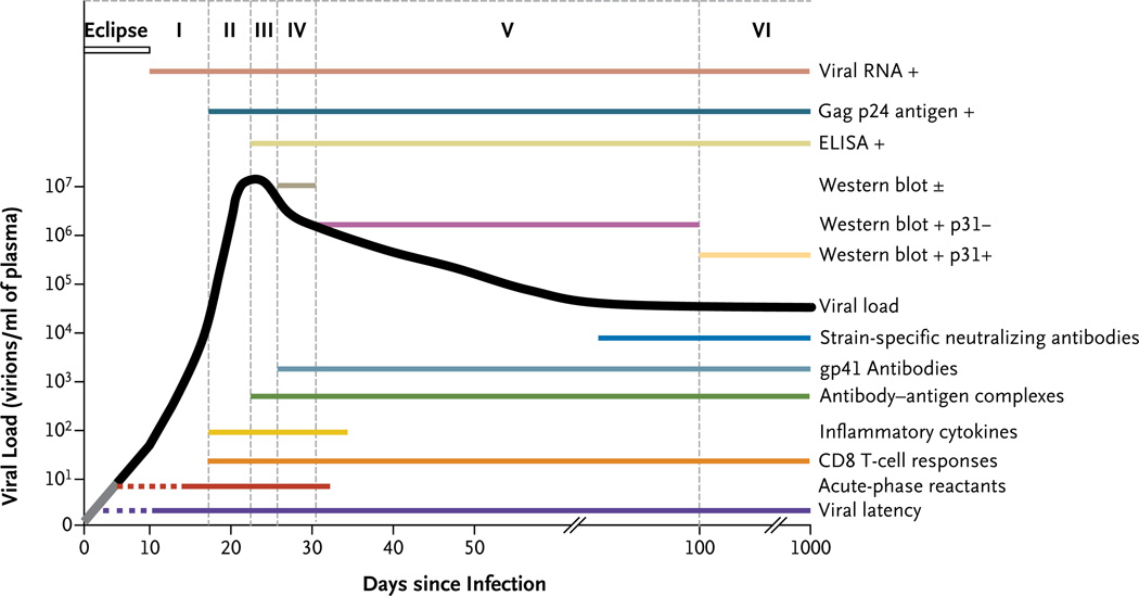
    1. Fiebig EW, Wright DJ, Rawal BD, et al. Dynamics of HIV viremia and antibody seroconversion in plasma donors: implications for diagnosis and staging of primary HIV infection. AIDS. 2003;17:1871–1879. - PubMed

Publication types