. 2011 May 19:10:59.
doi: 10.1186/1476-4598-10-59.

Targeting oncogenic miR-335 inhibits growth and invasion of malignant astrocytoma cells

Affiliations

Targeting oncogenic miR-335 inhibits growth and invasion of malignant astrocytoma cells

Minfeng Shu et al. Mol Cancer. .

Abstract

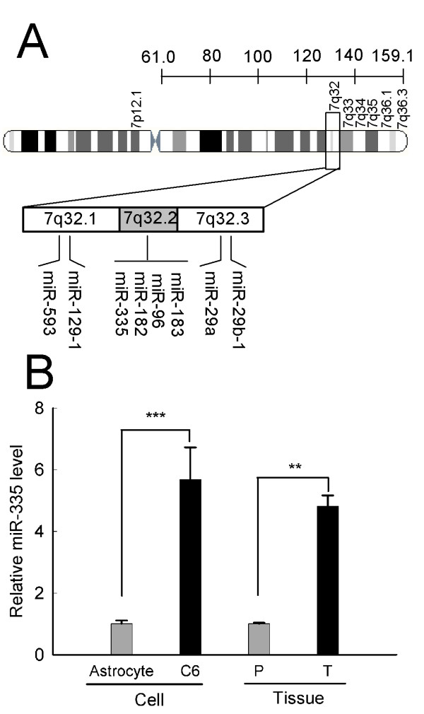
Background: Astrocytomas are the most common and aggressive brain tumors characterized by their highly invasive growth. Gain of chromosome 7 with a hot spot at 7q32 appears to be the most prominent aberration in astrocytoma. Previously reports have shown that microRNA-335 (miR-335) resided on chromosome 7q32 is deregulated in many cancers; however, the biological function of miR-335 in astrocytoma has yet to be elucidated.

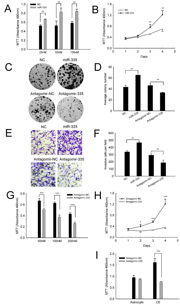
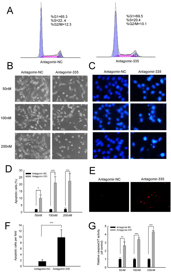
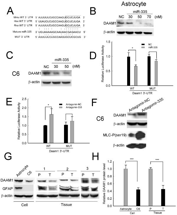
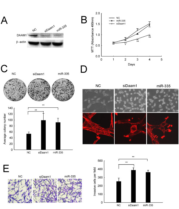
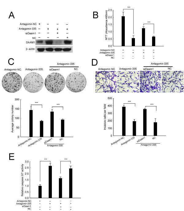
Results: We report that miR-335 acts as a tumor promoter in conferring tumorigenic features such as growth and invasion on malignant astrocytoma. The miR-335 level is highly elevated in C6 astrocytoma cells and human malignant astrocytomas. Ectopic expression of miR-335 in C6 cells dramatically enhances cell viability, colony-forming ability and invasiveness. Conversely, delivery of antagonist specific for miR-335 (antagomir-335) to C6 cells results in growth arrest, cell apoptosis, invasion repression and marked regression of astrocytoma xenografts. Further investigation reveals that miR-335 targets disheveled-associated activator of morphogenesis 1(Daam1) at posttranscriptional level. Moreover, silencing of endogenous Daam1 (siDaam1) could mimic the oncogenic effects of miR-335 and reverse the growth arrest, proapoptotic and invasion repression effects induced by antagomir-335. Notably, the oncogenic effects of miR-335 and siDAAM1 together with anti-tumor effects of antagomir-335 are also confirmed in human astrocytoma U87-MG cells.

Conclusion: These findings suggest an oncogenic role of miR-335 and shed new lights on the therapy of malignant astrocytomas by targeting miR-335.


Figures

Figure 1
Figure 1
MiR-335 is highly expressed in astrocytoma. (A) A schematic showing miRNAs located on chromosome 7q32 in astrocytoma. (B) Data shown are expression level of miR-335 in C6 astrocytoma cells and patient astrocytoma tissues by real-time quantitative RT-PCR, comparing with their relative normal counterparts. P, adjacent paracancerous tissues; T, patient astrocytoma tissues. RNA input was normalized by U6 snRNA. Data represent the means ± SD of three independent experiments. Statistical differences compared with the controls are given as **, P < 0.01; ***, P < 0.001.
Figure 2
Figure 2
Effects of miR-335 on viability and invasion in C6 astrocytoma cells. (A) Dose-dependent effect of miR-335 mimics on cell viability. C6 cells were transfected with miR-335 mimics or negative control mimics (NC) for 72 h at the indicated concentrations. (B) Time-dependent effect of miR-335 mimics on cell viability. C6 cells were transfected with 50 nM miR-335 mimics for the indicated times. (C, D) Colony formation was observed at the presence of miR-335 mimics (C up-panel) or antagomir-335(C down-panel). (E, F) Cell invasiveness was detected by transwell invasion assay. C6 cells were transfected with 50 nM miR-335 mimics (E up-panel) or 100 nM antagomir-335 (E down-panel) or their counterpart negative controls. Graph is the representative of three independent experiments. (G) Dose-dependent effect of antagomir-335 on cell viability. C6 cells were transfected with antagomir-335 or antagomir-NC for 72 h at the indicated concentrations. (H) Time-dependent effect of antagomir-335 on cell viability. C6 cells were transfected with 100 nM antagomir-335 for the indicated times. (I) Cell viability was measured with MTT assay. Rat normal astrocytes and C6 astrocytoma cells were transfected with 100 nM antagomir-335 or antagomir-NC for 72 h. Data represent the means ± SD. Statistical differences compared with the controls are given as *, P < 0.05; **, P < 0.01; ***, P < 0.001. Original magnification in (E), 200 ×.
Figure 3
Figure 3
MiR-335 abrogation induces apoptosis in C6 astrocytoma cells. (A) Cell cycle distribution detection. C6 cells were transfected with 100 nM antagomir-335 or antagomir-NC for 72 h and cell cycle distribution was analyzed by flow cytometer. (B) Phase-contrast imaging and (C) Hoechest 33258 staining of C6 cells. (D) Quantitation of the percentage of apoptotic cells. (E, F) Apoptotic cells were detected by TUNEL assay. (G) Dose-dependent effect of antagomir-335 on caspase3/7 activity. C6 cells were transfected with antagomir-335 at the indicated concentrations for 72 h. Data represent the means ± SD. Statistical differences compared with the controls are given as *, P < 0.05; **, P < 0.01; ***, P < 0.001. Original magnification in (B, E), 200 ×; Original magnification in (C), 640 ×.
Figure 4
Figure 4
Daam1 is a direct target of miR-335. (A) Schematic representation of 3'-UTR of Daam1 mRNA with the putative miR-335 binding sequence. Mutation was introduced to the Daam1 3'-UTR sequence in the complementary site for the seed region of miR-335. (B, C) Effects of miR-335 on expression of endogenous DAAM1 protein. Western blot analysis was used to monitor DAAM1 protein expression in rat astrocytes (B) and C6 cells (C) 72 h after transfection with miR-335 mimics or negative control mimics (NC) at the indicated concentrations. (D, E) Analysis of luciferase activity. C6 cells were cotransfected with psiCHECK-2-wild type Daam1 3'-UTR/-mutant Daam1 3'-UTR (indicated as WT or MUT on the × axis), and (D) miR-335 mimics or (E) antagomir-335, luciferase activity was assayed 72 h after transfection. (F) Effects of antagomir-335 on expression of DAAM1 and phosphorylation of MLC (myosin light chain). C6 cells were transfected with 100 nM antagomir-335 or antagomir-NC, and applied for western blot analysis 72 h later. (G) DAAM1 protein is significantly downregulated in astrocytoma. Endogenous DAAM1 expression in C6 cells and human astrocytoma tissues were detected by western blot. GFAP expression was used as a biomarker of normal astrocytes. (H) Quantified protein levels of DAAM1 are shown as normalized by β-actin. Data represent the means ± SD of three independent experiments. Statistical differences compared with the controls are given as *, P < 0.05; **, P < 0.01; ***, P < 0.001.
Figure 5
Figure 5
SiDaam1 mimics the oncogenic effects of miR-335 in C6 cells. (A) SiDaam1 efficiently inhibited the expression of DAAM1. Western blot was used to detect the expression of DAAM1 72 h after transfection with 50 nM siDaam1 or miR-335 mimics in C6 cells. (B) Cell viability was detected by MTT assay. (C) Effect of siDaam1 or miR-335 transfection on colony formation. (D) Morphologic alteration (up-panel) and phallotoxins stained actin rearrangement (down-panel). Cells transfected with siDaam1 or miR-335 adopted a stellate morphology and collapsed their actin stress fibers together with increased membrane ruffling. (E) Cell invasiveness was detected by transwell invasion assay. C6 cells were transfected with 50 nM siDaam1 or miR-335 mimics for the indicated times. Results represent the means ± SD for three repeats. (*, p < 0.05;**, p < 0.01). Original magnification in (D up-panel; E), 200 ×; (D down-panel), 630 ×.
Figure 6
Figure 6
SiDaam1 reverses the anti-tumor effects of antagomir-335 in C6 cells. (A) SiDaam1 obviously abrogated upregulation of DAAM1 induced by antagomir-335 transfection. (B-E) siDaam1 counteracted the anti-tumor effects of antagomir-335. SiDaam1 significantly abrogated antagomir-335-induced growth arrest (B), repression of colony formation(C), invasion inhibition (D) and caspase3/7 activity (E). C6 cells were transfected with 50 nM siDaam1 and/or 100 nM antagomir-335 for the indicated times. Results represent the means ± SD for three repeats. (***, p < 0.001). Original magnification in (D), 200 ×.
Figure 7
Figure 7
MiR-335 inhibition suppresses tumor growth in vivo. (A-C) Pretreated effect of antagomir-335 on tumor formation in nude mouse xenograft model. (A)Antagomir-NC-transfected and antagomir-335-transfected C6 cells were s.c. injected into the left (up-panel) and right (down-panel) flanks of nude mice, respectively (n = 8). (B) Tumor volume was monitored during the time course of 19 days. (C) Tumor weight was detected at the end of the study. (D-H) Therapeutic effect of antagomir-335 on tumor growth in nude mouse xenograft model. (D) Tumors generated by C6 cells injection in nude mice were intratumorally injected with antagomir-NC (up-panel) and antagomir-335 (down-panel) for 2 weeks, respectively(n = 5). (E) Tumor volume was monitored during the time course of 4 weeks. (F) Tumor weight was detected at the end of the study. (G) DAAM1 expression was detected by immunohistochemistry. (H) Apoptotic cells were detected by TUNEL assay. Data represent the means ± SD. Statistical differences compared with the controls are given as **, P < 0.01; ***, P < 0.001. Original magnification in (G up-panel), 320 ×; Original magnification in (H), 400 ×.
Figure 8
Figure 8
Both siDAAM1 and miR-335 promote growth and invasion of human astrocytoma U87-MG cells. (A) miR-335 expression in U87-MG and HEB cells was detected by real-time qRT-PCR. (B) Cell viability was detected by MTT assay. (C) Effect of siDAAM1 or miR-335 transfection on colony formation. (D) Effect of siDAAM1 transfection on DAAM1 protein expression. U87-MG cells were transfected with 50 nM siDAAM1 for 72 h. (E) Morphologic alteration (up-panel) and phallotoxins stained actin rearrangement (down-panel). Cells treated with siDAAM1 or miR-335 adopted a stellate morphology and collapsed their actin stress fibers together with increased membrane ruffling. (F) Cell invasiveness was detected by transwell invasion assay. (G left-panel) Western blot analysis was used to detect DAAM1 protein expression in U87-MG cells 72 h after transfection with 100 nM miR-335 mimics. (G right-panel) Endogenous DAAM1 expression in U87-MG and HEB cells was detected by western blot. GFAP expression was used as a biomarker of normal astrocytes. Results represent the means ± SD for three repeats. (*, p < 0.05;**, p < 0.01;***, P < 0.001). Original magnification in (E up-panel; F), 200 ×; (E down-panel), 400 ×.
Figure 9
Figure 9
MiR-335 inhibition induces apoptosis and suppresses invasion of human astrocytoma U87-MG cells. (A) Time-dependent effect of antagomir-335 on cell viability. U87-MG cells were transfected with 100 nM antagomir-335 for the indicated times. (B) Effect of antagomir-335 transfection on colony formation. (C) Cell invasiveness was detected by transwell invasion assay. U87-MG cells were transfected with 100 nM antagomir-335 or antagomir-NC. Graph is the representative of three independent experiments. (D) Phase-contrast imaging and (E) Hoechest 33258 staining of U87-MG cells. (F) Apoptotic cells were detected by TUNEL assay. (G) Effect of antagomir-335 on caspase3/7 activity. U87-MG cells were transfected with 100 nM antagomir-335 or antagomir-NC for 72 h. (H) Effect of antagomir-335 on expression of DAAM1 (left-panel) and phosphorylation of MLC (right-panel) in U87-MG cells. Cells were transfected with 100 nM antagomir-335 or antagomir-NC for 48 h. Data represent the means ± SD. Statistical differences compared with the controls are given as *, P < 0.05; **, p < 0.01; ***, P < 0.001. Original magnification in (C, D, F), 200 ×; Original magnification in (E), 400 ×.

References

    1. DeAngelis LM. Brain tumors. N Engl J Med. 2001;344:114–123. doi: 10.1056/NEJM200101113440207. - DOI - PubMed
    1. Gabriely G, Wurdinger T, Kesari S, Esau CC, Burchard J, Linsley PS, Krichevsky AM. MicroRNA 21 promotes glioma invasion by targeting matrix metalloproteinase regulators. Mol Cell Biol. 2008;28:5369–5380. doi: 10.1128/MCB.00479-08. - DOI - PMC - PubMed
    1. Esquela-Kerscher A, Slack FJ. Oncomirs-microRNAs with a role in cancer. Nat Rev Cancer. 2006;6:259–269. doi: 10.1038/nrc1840. - DOI - PubMed
    1. Medina PP, Slack FJ. microRNAs and cancer: an overview. Cell Cycle. 2008;7:2485–2492. doi: 10.4161/cc.7.16.6453. - DOI - PubMed
    1. Ventura A, Jacks T. MicroRNAs and cancer: short RNAs go a long way. Cell. 2009;136:586–591. doi: 10.1016/j.cell.2009.02.005. - DOI - PMC - PubMed

Publication types

MeSH terms

LinkOut - more resources