

Association between a quantitative CT scan measure of brain edema and outcome after cardiac arrest
- PMID: 21592642
- PMCID: PMC3244968
- DOI: 10.1016/j.resuscitation.2011.04.001
Association between a quantitative CT scan measure of brain edema and outcome after cardiac arrest
Abstract
Background: Cerebral edema is one physical change associated with brain injury and decreased survival after cardiac arrest. Edema appears on computed tomography (CT) scan of the brain as decreased X-ray attenuation by gray matter. This study tested whether the gray matter attenuation to white matter attenuation ratio (GWR) was associated with survival and functional recovery.
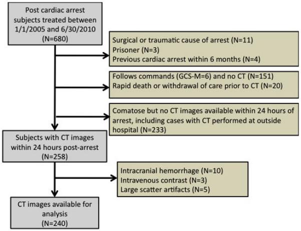
Methods: Subjects were patients hospitalized after cardiac arrest at a single institution between 1/1/2005 and 7/30/2010. Subjects were included if they had non-traumatic cardiac arrest and a non-contrast CT scan within 24h after cardiac arrest. Attenuation (Hounsfield Units) was measured in gray matter (caudate nucleus, putamen, thalamus, and cortex) and in white matter (internal capsule, corpus callosum and centrum semiovale). The GWR was calculated for basal ganglia and cerebrum. Outcomes included survival and functional status at hospital discharge.
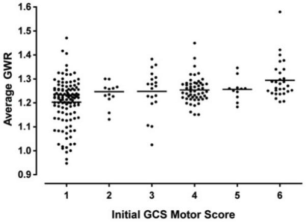
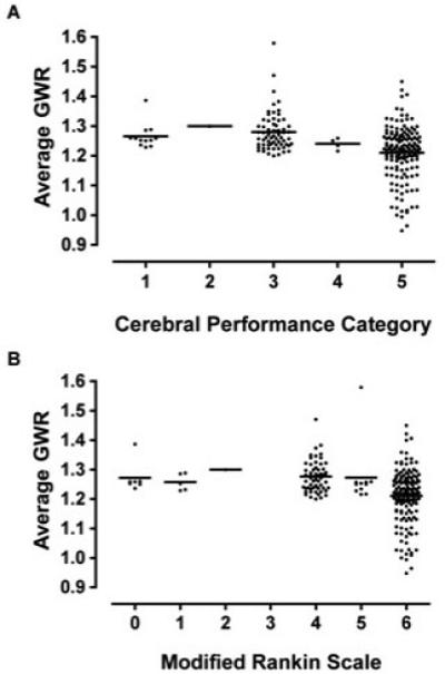
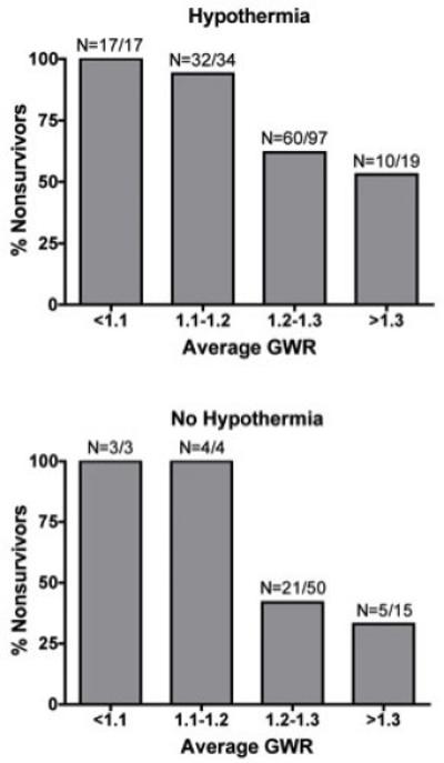
Results: For 680 patients, 258 CT scans were available, but 18 were excluded because of hemorrhage (10), intravenous contrast (3) or technical artifact (5), leaving 240 CT scans for analysis. Lower GWR values were associated with lower initial Glasgow Coma Scale motor score. Overall survival was 36%, but decreased with decreasing GWR. The average of basal ganglia and cerebrum GWR provided the best discrimination. Only 2/58 subjects with average GWR<1.20 survived and both were treated with hypothermia. The association of GWR with functional outcome was completely explained by mortality when GWR<1.20.
Conclusions: Subjects with severe cerebral edema, defined by GWR<1.20, have very low survival with conventional care, including hypothermia. GWR estimates pre-treatment likelihood of survival after cardiac arrest.
Copyright © 2011 Elsevier Ireland Ltd. All rights reserved.
Figures
Comment in
-
Imaging brain injury after cardiac arrest resuscitation when it really matters.Resuscitation. 2011 Sep;82(9):1124-5. doi: 10.1016/j.resuscitation.2011.05.029. Epub 2011 Jun 14. Resuscitation. 2011. PMID: 21719185 No abstract available.
References
-
- Neumar RW, Nolan JP, Adrie C, Aibiki M, Berg RA, Böttiger BW, Callaway C, Clark RS, Geocadin RG, Jauch EC, Kern KB, Laurent I, Longstreth WT, Jr, Merchant RM, Morley P, Morrison LJ, Nadkarni V, Peberdy MA, Rivers EP, Rodriguez-Nunez A, Sellke FW, Spaulding C, Sunde K, Vanden Hoek T. Post-cardiac arrest syndrome: epidemiology, pathophysiology, treatment, and prognostication. Circulation. 2008;118:2452–2483. - PubMed
-
- Laver S, Farrow C, Turner D, Nolan J. Mode of death after admission to an intensive care unit following cardiac arrest. Intensive Care Med. 2004;30:2126–2128. - PubMed
-
- van Alem AP, Waalewijn RA, Koster RW, de Vos R. Assessment of quality of life and cognitive function after out-of-hospital cardiac arrest with successful resuscitation. Am J Cardiol. 2004;93:131–135. - PubMed
-
- Pusswald G, Fertl E, Faltl M, Auff E. Neurological rehabilitation of severely disabled cardiac arrest survivors. Part II. Life situation of patients and families after treatment. Resuscitation. 2000;47:241–248. - PubMed
Publication types
MeSH terms
Grants and funding
LinkOut - more resources
Full Text Sources
Other Literature Sources
Medical
