Review
. 2011 Dec;22(6):901-8.
doi: 10.1016/j.copbio.2011.04.020. Epub 2011 May 16.

Applications of viral nanoparticles in medicine

Affiliations
Review

Applications of viral nanoparticles in medicine

Ibrahim Yildiz et al. Curr Opin Biotechnol. 2011 Dec.

Abstract

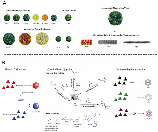
Several nanoparticle platforms are currently being developed for applications in medicine, including both synthetic materials and naturally occurring bionanomaterials such as viral nanoparticles (VNPs) and their genome-free counterparts, virus-like particles (VLPs). A broad range of genetic and chemical engineering methods have been established that allow VNP/VLP formulations to carry large payloads of imaging reagents or drugs. Furthermore, targeted VNPs and VLPs can be generated by including peptide ligands on the particle surface. In this article, we highlight state-of-the-art virus engineering principles and discuss recent advances that bring potential biomedical applications a step closer. Viral nanotechnology has now come of age and it will not be long before these formulations assume a prominent role in the clinic.


Figures

Figure 1
Figure 1
A. Viral nanoparticles (VNPs) used in materials science and medicine. Icosahedral plant viruses: Brome mosaic virus (BMV), Cowpea cholorotic mottle virus (CCMV), Cowpea mosaic virus (CPMV), Hibiscus cholorotic ringspot virus (HCRSV), Red clover necrotic mottle virus (RCNMV), Turnip yellow mosaic virus (TYMV). Icosahdral insect virus: Flock House virus (FHV). Icosahedral bacteriophages: HK97, P22, T7, MS2 and Qβ. Note that P22 and T7 are head-tail phages, with the tails not shown. Icosahedral mammalian virus: Adenovirus (Ad). Rod-shaped and filamentous viruses: Potato virus X (PVX), Tobacco mosaic virus (TMV), bacteriophage M13. Images of the following VNPs were reproduced from the VIPER Database; URL: http://www.viperdb.scripps.edu/: BMV, CCMV, CPMV, P22, TYMV, FHV, HK97, MS2, Ad, and Qβ. The structures of HCRSV, RCNMV, T7, PVX and TMV were reproduced from refs [–18], respectively. B. Genetic, chemical, and self-assembly/encapsulation manipulations of VNPs in biomedical research.
Figure 2
Figure 2
Intravital imaging of viral nanoparticle uptake in prostate tumors in vivo. A. Intravital fluorescence confocal imaging of PC-3 prostate tumor (green channel) showing uptake of AF647-labeled CPMV-PEG-bombesin (heat map) over time. Images are representative of n=10 experiments. Colors correspond to tumor/stroma ratio (see key). Scale bar = 3 mm. B. Intravital imaging of PC-3 prostate tumor (green channel) showing uptake of AF647-labeled CPMV-PEG (heat map) over time. Images are representative of n=10 experiments. Colors correspond to tumor/stroma ratio (see key). Scale bar = 3 mm. C. Quantitation of tumor uptake of CPMV conjugates over time, n=10 experiments per group. Values expressed as mean tumor/stroma ratio, using GFP channel to delineate tumor. Uptake of CPMV-PEG-bombesin is significantly higher than CPMV-PEG at 2 h and beyond (P<0.0001). D. Accumulation of CPMV conjugates in tumor tissue, measured by fluorescence confocal microscopy of tissue sections. Grayscale and color merged images are provided with PC-3 GFP cells (green) and CPMV-AF647 conjugates (red). Scale bar = 75 μm. Reproduced from Ref .
Figure 3
Figure 3
Volume-rendered PET images of male Sprague–Dawley rats injected with [18F]-MS2. Sprague–Dawley rats were anesthetized and placed side-by-side in a PET scanner. The animals were injected at the same time through the tail vein with same amount of either of [18F]-fluorobenzaldehyde or [18F]-MS2. [18F]-fluorobenzaldehyde was rapidly cleared from circulation, with essentially no signal in the heart blood pool after only 15 seconds. In contrast, [18F]-MS2 remained in circulation for the duration of the experiment (3 hours) as seen in the figure. Reproduced with permission from reference [76].

References

    1. Resch-Genger U, Grabolle M, Cavaliere-Jaricot S, Nitschke R, Nann T. Quantum dots versus organic dyes as fluorescent labels. Nat Methods. 2008;5(9):763–775. - PubMed
    1. McNerny DQ, Leroueil PR, Baker JR. Understanding specific and nonspecific toxicities. a requirement for the development of dendrimer-based pharmaceuticals. Wires Nanomed Nanobi. 2010;2(3):249–259. - PMC - PubMed
    1. Jesorka A, Orwar O, Liposomes Technologies and Analytical Applications. Annu Rev Anal Chem. 2008;1:801–832. - PubMed
    1. Giljohann DA, Seferos DS, Daniel WL, Massich MD, Patel PC, Mirkin CA. Gold Nanoparticles for Biology and Medicine. Angew Chem Int Edit. 2010;49 (19):3280–3294. - PMC - PubMed
    1. Fortin JP, Wilhelm C, Servais J, Menager C, Bacri JC, Gazeau F. Size-sorted anionic iron oxide nanomagnets as colloidal mediators for magnetic hyperthermia. J Am Chem Soc. 2007;129(9):2628–2635. - PubMed

Publication types