. 2011 Jul 15;286(28):25016-26.
doi: 10.1074/jbc.M111.246637. Epub 2011 May 18.

Antibody recognition of a human chorionic gonadotropin epitope (hCGbeta66-80) depends on local structure retained in the free peptide

Affiliations

Antibody recognition of a human chorionic gonadotropin epitope (hCGbeta66-80) depends on local structure retained in the free peptide

Craig R Gregor et al. J Biol Chem. .

Abstract

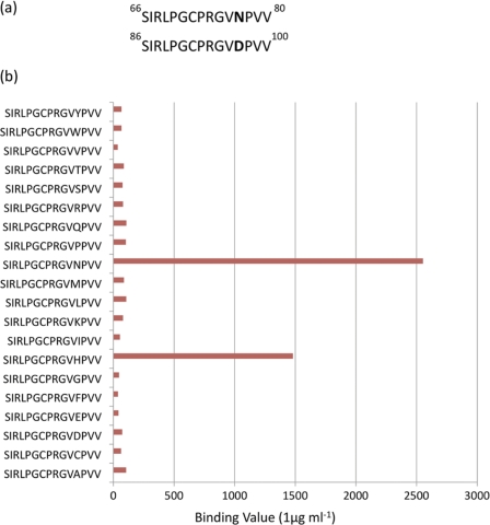
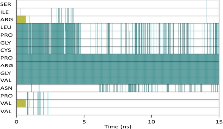
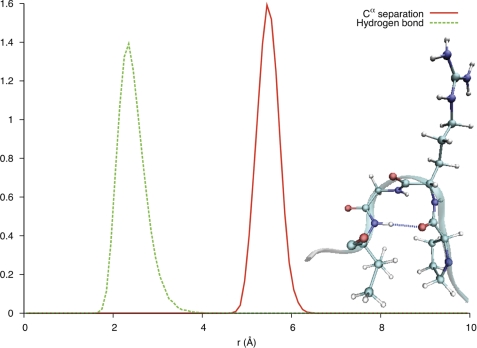
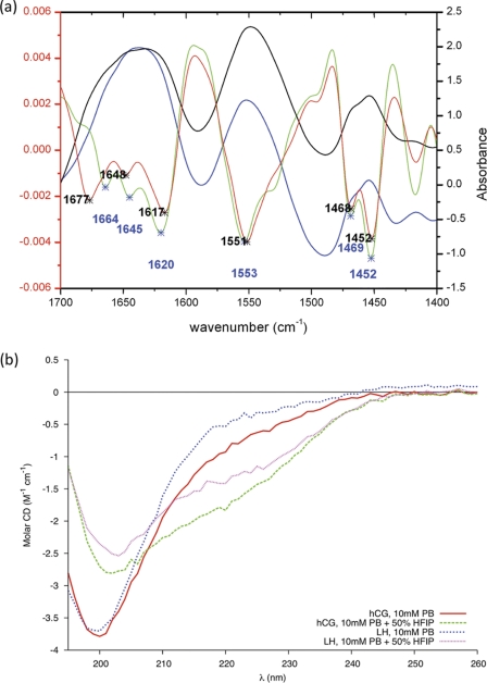
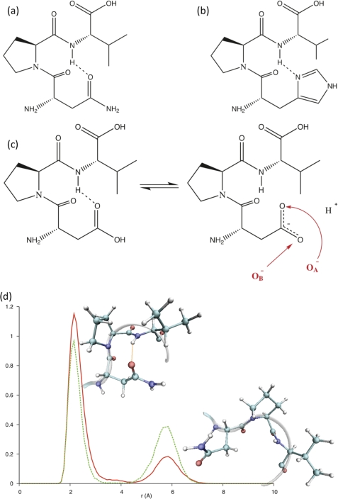
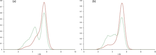
Human chorionic gonadotropin (hCG) is an important biomarker in pregnancy and oncology, where it is routinely detected and quantified by specific immunoassays. Intelligent epitope selection is essential to achieving the required assay performance. We present binding affinity measurements demonstrating that a typical β3-loop-specific monoclonal antibody (8G5) is highly selective in competitive immunoassays and distinguishes between hCGβ(66-80) and the closely related luteinizing hormone (LH) fragment LHβ(86-100), which differ only by a single amino acid residue. A combination of optical spectroscopic measurements and atomistic computer simulations on these free peptides reveals differences in turn type stabilized by specific hydrogen bonding motifs. We propose that these structural differences are the basis for the observed selectivity in the full protein.


Figures

FIGURE 1.
FIGURE 1.
a, crystal structure of hCG (1). hCGβ66–80 is circled. b, crystal structure of hCGβ66–80 as obtained from Protein Data Bank ID code 1HRP. Hydrogen atoms are omitted.
FIGURE 2.
FIGURE 2.
a, primary structure of hCGβ66–80 (upper) and LHβ86–100 (lower). b, binding results from a small subset of the Pepscan positional substitution library, in which there is a systematic substitution of position 77, occupied by asparagine in hCGβ. Note that there is negligible binding to the peptide occupying the position third from the bottom, which equates to the sequence in LH. In the sequence containing histidine at position 77, the binding is ∼60% that of the hCGβ binding (supplemental Table 1).
FIGURE 3.
FIGURE 3.
Secondary structure of each residue of hCGβ66–80 as a function of time. Green corresponds to turn, white is unordered, and gold is β-strand. Simulation predicts a stable turn over Pro73–Val76, whereas a second turn spanning Leu69–Cys72 was found to be only a transient feature.
FIGURE 4.
FIGURE 4.
Solid red line, separation of Cα in Pro73 and Val76. Dashed green line, separation between backbone carbonyl (Pro73) and amine (Val76) groups. Inset, visualization of the turn motif spanning residues PRGV (Pro73–Val76), revealing the stabilizing hydrogen bond between the backbone carbonyl group of Pro73 and the backbone amine group of Val76.
FIGURE 5.
FIGURE 5.
a, FTIR-attenuated total reflectance blank-subtracted and TFA-corrected spectrum of hCGβ66–80 (black), LHβ86–100 (blue), and their second derivative spectra (red and green, respectively) displaying the peak positions of the different components. b, far-ultraviolet CD spectrum of 0.11 mm hCGβ66–80 and 0.11 mm LHβ86–100.
FIGURE 6.
FIGURE 6.
Secondary structure of each residue of LHβ86–100 as a function of time. Green corresponds to turn, white is unordered, and gold is β-strand. Simulation predicts a stable turnover Pro93–Val96, with the span extending to include Asp97 and Pro98. A second turn spanning Leu89–Cys92 was found to be less stable.
FIGURE 7.
FIGURE 7.
Solid red line, Cα separation. Dashed green line, separation between backbone carbonyl and amine. a, Pro93–Val96. b, Asp97–Val100.
FIGURE 8.
FIGURE 8.
Ramachandran angles of sequence PRGV in hCGβ66–80 (a) and LHβ86–100 (b) over the last 5 ns. The torsional angles of the two central amino acids, Arg74 and Gly75, indicate a type I β-turn, whereas those for Arg94 and Gly95 indicate a type II β-turn.
FIGURE 9.
FIGURE 9.
a–c, proposed H-bonding geometry in hCGβ66–80 (a), His77 mutant (b), and Asp77 mutant depicting the proposed “clasp” being only present in the protonated form of Asp (c). d, bond probability analysis of the safety catch in hCGβ66–80, coupled with visualizations of the structure corresponding to each peak.
FIGURE 10.
FIGURE 10.
Separation of Asp97 carboxylate oxygen (a) O−A, (b) O−B, and hydrogen of the backbone amine group of Val79. Solid red line, over full 15 ns. Dashed green line, over last 5 ns.

Similar articles

Cited by

References

    1. Lapthorn A. J., Harris D. C., Littlejohn A., Lustbader J. W., Canfield R. E., Machin K. J., Morgan F. J., Isaacs N. W. (1994) Nature 369, 455–461 - PubMed
    1. Cole L. A. (2007) Placenta 28, 977–986 - PubMed
    1. O'Connor J. F., Birken S., Lustbader J. W., Krichevsky A., Chen Y., Canfield R. E. (1994) Endocr. Rev. 15, 650–683 - PubMed
    1. Sturgeon C. M., McAllister E. J. (1998) Ann. Clin. Biochem. 35, 460–491 - PubMed
    1. Gronowski A. M., Grenache D. G. (2009) Clin. Chem. 55, 1447–1449 - PubMed

Publication types

Substances

LinkOut - more resources