. 2011 Jul 22;286(29):25770-7.
doi: 10.1074/jbc.M110.204503. Epub 2011 May 18.

Integrin βν-mediated phagocytosis of apoptotic cells in Drosophila embryos

Affiliations

Integrin βν-mediated phagocytosis of apoptotic cells in Drosophila embryos

Kaz Nagaosa et al. J Biol Chem. .

Abstract

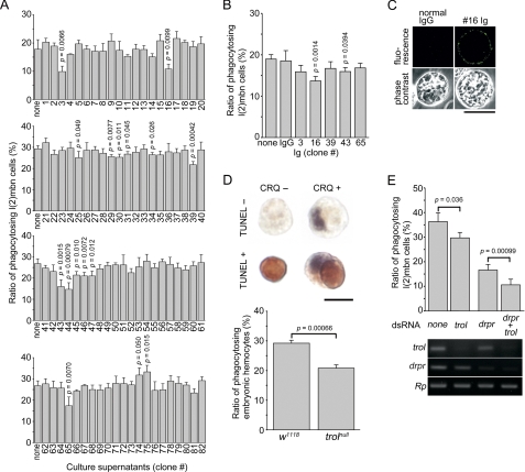
To identify molecules that play roles in the clearance of apoptotic cells by Drosophila phagocytes, we examined a series of monoclonal antibodies raised against larval hemocytes for effects on phagocytosis in vitro. One antibody that inhibited phagocytosis recognized terribly reduced optic lobes (Trol), a core protein of the perlecan-type proteoglycan, and the level of phagocytosis in embryos of a Trol-lacking fly line was lower than in a control line. The treatment of a hemocyte cell line with a recombinant Trol protein containing the amino acid sequence RGD augmented the phosphorylation of focal adhesion kinase, a hallmark of integrin activation. A loss of integrin βν, one of the two β subunits of Drosophila integrin, brought about a reduction in the level of apoptotic cell clearance in embryos. The presence of integrin βν at the surface of embryonic hemocytes was confirmed, and forced expression of integrin βν in hemocytes of an integrin βν-lacking fly line recovered the defective phenotype of phagocytosis. Finally, the level of phagocytosis in a fly line that lacks both integrin βν and Draper, another receptor required for the phagocytosis of apoptotic cells, was lower than that in a fly line lacking either protein. We suggest that integrin βν serves as a phagocytosis receptor responsible for the clearance of apoptotic cells in Drosophila, independent of Draper.


Figures

FIGURE 1.
FIGURE 1.
Identification of Trol that plays a role in the phagocytosis of apoptotic cells by Drosophila hemocytes. A, an assay for phagocytosis was conducted with ecdysone-treated l(2)mbn cells as phagocytes and apoptotic S2 cells as targets in the presence and absence of culture supernatants of the indicated hybridoma clones producing monoclonal antibodies raised against larval hemocytes. The ratio of l(2)mbn cells that had accomplished phagocytosis is shown. Significance is versus none. Data from one experiment are presented (S.D. are from data with four microscopic fields). B, the same assay as in A was conducted in the presence of immunoglobulin prepared from culture supernatants of the indicated hybridoma clones or normal mouse IgG (2 μg/ml). The ratio of l(2)mbn cells that had accomplished phagocytosis is shown. Significance is versus control IgG. Data from one of two independent experiments with similar results are presented (S.D. are from data with six microscopic fields). C, ecdysone-treated l(2)mbn cells were subjected to immunocytochemistry with the #16 immunoglobulin or normal rat IgG as a negative control under membrane-nonpermeabilizing conditions. Phase-contrast and fluorescence micrographs of the same fields are shown. Scale bar, 20 μm. D, phagocytosis of apoptotic cells was examined with dispersed embryonic cells of the indicated fly lines. Embryonic cells were cytochemically analyzed with an anti-Croquemort (CRQ) antibody (signals seen in purple) and by TUNEL (signals seen in brown) (top). Scale bar, 5 μm. The ratio of hemocytes that had accomplished phagocytosis is shown (bottom). The genotype of trolnull is trolnull FRT101/Y. E, l(2)mbn cells, which had been treated with double-stranded RNA (dsRNA) containing sequences of mRNA of the indicated genes, were analyzed for the phagocytosis of apoptotic S2 cells as in A (top) as well as for the level of mRNA of the indicated genes by RT-PCR (bottom). In the top panel, data from one of three independent experiments with similar results are presented (S.D. are from data with six microscopic fields). drpr, draper; Rp, gene coding for ribosomal protein S15Ab.
FIGURE 2.
FIGURE 2.
Identification of integrin βν required for the phagocytosis of apoptotic cells in Drosophila embryos. A, the structure of Trol (a product from transcript variant C) is schematically exhibited with the positions of the motif RGD. Shown below are portions of Trol prepared as recombinant proteins fused to GST (GST-Trol 1 and GST-Trol 2). The numbers are amino acid positions with the amino terminus numbered 1. B, top, l(2)mbn cells that had been incubated with the indicated proteins were immunocytochemically analyzed for the bound proteins using anti-GST antibody. Phase-contrast and fluorescence micrographs of the same fields are shown. The arrowheads point to the positive signals. Scale bar, 10 μm. Bottom, lysates (20 μg of protein) of l(2)mbn cells that had been incubated in culture containers coated with the indicated proteins were analyzed by Western blotting with the anti-phospho-FAK antibody and anti-FAk56 antibody. The arrowheads indicate the positions of phosphorylated FAK56 and total FAK56. The intensity of the signals was determined, and averages from three independent experiments are shown relative to the result with a negative control (GST). C, lysates (0.13 mg of protein) of larvae of the indicated fly lines were analyzed by Western blotting with the anti-integrin βν antibody. The arrowhead indicates the position of integrin βν. D, dispersed embryonic cells of the indicated fly lines were analyzed for the phagocytosis of apoptotic cells by hemocytes (top), the population of hemocytes (middle), and the number of apoptotic cells (bottom). The ratios in percentage terms of hemocytes that had accomplished phagocytosis (top), of hemocytes to all embryonic cells (middle), and of apoptotic cells to all embryonic cells (bottom) are shown. Genotypes of the fly lines analyzed are w; betaInt-nu1 (betaInt-nu1), w; betaInt-nu2 (betaInt-nu2), mys1/Y (mys1), and sn mysXG43/Y (mysXG43). Significance is versus w1118.
FIGURE 3.
FIGURE 3.
Presence and function of integrin βν in hemocytes. A, lysates (0.1 mg of protein) of w1118 flies at the indicated developmental stages were analyzed by Western blotting with the anti-integrin βν antibody. The arrowhead points to the position of integrin βν. B, l(2)mbn cells (left) and dispersed embryonic cells of y w; srpHemo-GAL4 UAS-srcEGFP flies (right) were subjected to immunocytochemistry with the anti-integrin βν antibody or preimmune rat serum as a negative control under membrane-nonpermeabilizing conditions. Embryonic hemocytes were identified based on the expression of GFP driven by the promoter of serpent. Note that most GFP-positive cells were also positive for the expression of Croquemort (data not shown). Phase-contrast and fluorescence micrographs of the same fields are shown. The arrowheads point to the signals derived from the anti-integrin βν antibody. Scale bars, 10 μm. C, dispersed embryonic cells of the indicated flies were analyzed for the phagocytosis of apoptotic cells by hemocytes. The ratio of hemocytes that had accomplished phagocytosis to total hemocytes is shown. Genotypes of the fly lines analyzed are w; betaInt-nu2 (betaInt-nu2 GAL4− UAS−), w; betaInt-nu2/betaInt-nu2 srpHemoGAL4 UAS-srcEGFP (betaInt-nu2 GAL4+ UAS−), w; betaInt-nu2; UAS-betaInt-nu/+ (betaInt-nu2 GAL4− UAS+), and w; betaInt-nu2/betaInt-nu2 srpHemoGAL4 UAS-srcEGFP; UAS-betaInt-nu/+ (betaInt-nu2 GAL4+ UAS+).
FIGURE 4.
FIGURE 4.
Independent actions of integrin βν and Draper. A, dispersed embryonic cells of the indicated flies were analyzed for the phagocytosis of apoptotic cells by hemocytes. The ratio of hemocytes that had accomplished phagocytosis to total hemocytes is shown. B, periods in the development of Drosophila from embryos to adults were determined with the indicated fly lines. Genotypes of the fly lines analyzed are w; betaInt-nu2 (betaInt-nu2), w; +; drprΔ5 (drprΔ5), and w; betaInt-nu2; drprΔ5 (betaInt-nu2 drprΔ5).

Similar articles

Cited by

References

    1. Savill J., Fadok V. (2000) Nature 407, 784–788 - PubMed
    1. Liao D. J. (2005) Med. Hypotheses 65, 23–28 - PubMed
    1. Nakanishi Y., Nagaosa K., Shiratsuchi A. (2011) Dev. Growth Differ. 53, 149–160 - PubMed
    1. Lauber K., Blumenthal S. G., Waibel M., Wesselborg S. (2004) Mol. Cell 14, 277–287 - PubMed
    1. Ravichandran K. S., Lorenz U. (2007) Nat. Rev. Immunol. 7, 964–974 - PubMed

Publication types

MeSH terms