. 2011 Jun 1;203(11):1574-81.
doi: 10.1093/infdis/jir084.

Fully human monoclonal antibody directed to proteolytic cleavage site in severe acute respiratory syndrome (SARS) coronavirus S protein neutralizes the virus in a rhesus macaque SARS model

Affiliations

Fully human monoclonal antibody directed to proteolytic cleavage site in severe acute respiratory syndrome (SARS) coronavirus S protein neutralizes the virus in a rhesus macaque SARS model

Tohru Miyoshi-Akiyama et al. J Infect Dis. .

Abstract

Background: There is still no effective method to prevent or treat severe acute respiratory syndrome (SARS), which is caused by SARS coronavirus (CoV). In the present study, we evaluated the efficacy of a fully human monoclonal antibody capable of neutralizing SARS-CoV in vitro in a Rhesus macaque model of SARS.

Methods: The antibody 5H10 was obtained by vaccination of KM mice bearing human immunoglobulin genes with Escherichia coli-producing recombinant peptide containing the dominant epitope of the viral spike protein found in convalescent serum samples from patients with SARS.

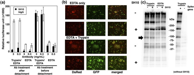
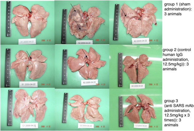
Results: 5H10, which recognized the same epitope that is also a cleavage site critical for the entry of SARS-CoV into host cells, inhibited propagation of the virus and pathological changes found in Rhesus macaques infected with the virus through the nasal route. In addition, we analyzed the mode of action of 5H10, and the results suggested that 5H10 inhibited fusion between the virus envelope and host cell membrane. 5H10 has potential for use in prevention and treatment of SARS if it reemerges.

Conclusions: This study represents a platform to produce fully human antibodies against emerging infectious diseases in a timely and safe manner.


Figures

Figure 1.
Figure 1.
Inhibitory effects of 5H10 on cell–cell fusion mediated by S-ACE2 interaction. A, Effects of 5H10 in cell–cell fusion analysis using a dual-reporter system. Fusion of S-expressing 293 cells and human ACE2-expressing 293 cells was quantified as described in Materials and Methods. Cells were detached with or without trypsin in addition to EDTA. The antibody treatments were performed before or after detachment as indicated. B, Effects of 5H10 on syncytium formation by S-expressing cells and human ACE2-expressing cells. Syncytium formation of S- and ACE2-expressing cells labeled with GFP and DsRed, respectively, was visualized by confocal laser microscopy. Cells were detached with or without trypsin in addition to EDTA, then mixed, and 5H10 was added to the cultures as indicated. C, Western blotting analysis to determine the effects of 5H10 on S cleavage by trypsin treatment. S-expressing 293T cells were treated with trypsin or EDTA in the presence or absence of 5H10 (3 mg/mL) as indicated. After lysis of the cells, Western blotting using anti-S antibody mixture was performed. Control 293T cells, which did not harbor the spike gene, were used as a negative control.
Figure 2.
Figure 2.
Gross pathological characterization of the lung damage in animals infected with SARS-CoV. Three animals were grouped as indicated and received sham treatment (top), control human IgG (middle), or 5H10 (bottom). Pathological changes found in the organs are indicated by arrows.
Figure 3.
Figure 3.
Viral load in lung tissues of animals administered 5H10. (a) SARS-CoV load was quantified by real-time RT-PCR. Data are presented as means ± SEM. (b) SARS-CoV genome fragments were amplified by RT-PCR. Data from each animal are presented and GAPDH was used to normalize the amounts of samples.
Figure 4.
Figure 4.
Immunohistochemical (A) and pathological (B) analyses of lung tissues from sham administration controls (a), those administered control human IgG (b), or those administered 5H10 (c). (A) Lung sections were treated with a mouse monoclonal antibody against SARS-CoV tissue sections were treated with the anti-SARS-CoV antibody, HRP-conjugated antibody, and DAB mix serially and stained with hematoxylin. (B) Histopathological analysis. (a) Sham administration group. Animal 1, +++, typical symptoms of acute DAD, extensive exudation and septal broadening, shrinkage of alveoli caused by pressure, restricted fusion of thick septa, ruptured elastic fibers of the alveoli, variably filled with protein-rich edema fluid, fibrin, erythrocytes, cellular debris, and a moderate number of inflammatory cells in alveolar cavities. Animal 2, ++++ severe acute DAD, massive cell infiltration and alveolar shrinkage, sheets of septal fusion, necrotic lesions at the hemorrhagic septa, and massive cell infiltration in alveolar cavities. Animal 3, ++++ severe acute DAD, massive cell infiltration and alveolar shrinkage, sheets of septal fusion, necrotic lesions at the hemorrhagic septa, and massive cell infiltration in alveolar cavities. (b) hIgG administration group. Animal 1, +++, typical symptoms of acute DAD, extensive exudation and septal broadening, shrinkage of alveoli caused by pressure, restricted fusion of thick septa, ruptured elastic fibers of alveoli, variably filled with protein-rich edema fluid, fibrin, erythrocytes, cellular debris, and a moderate number of inflammatory cells in alveolar cavities. Animal 2, +++, typical symptoms of acute DAD, extensive exudation and septal broadening, shrinkage of alveoli caused by pressure, restricted fusion of thickened septa, ruptured elastic fibers of alveoli, variably filled with protein-rich edema fluid, fibrin, erythrocytes, cellular debris, and a moderate number of inflammatory cells in alveolar cavities. Animal 3, ++ early symptoms of acute DAD, alveolar septal broadening with increasing inflammatory cell infiltration. (c) 5H10 administration group. Animal 1, + apparent inflammation, hemorrhage in septa, elastic fibers of alveolar wall distorted as shown by silver staining. Animal 2, ++ early symptoms of acute DAD, alveolar septal broadening with increasing inflammatory cell infiltration. Animal 3, ++ early symptoms of acute DAD, alveolar septal broadening with increasing inflammatory cell infiltration.

Similar articles

Cited by

References

    1. Guan Y, Zheng BJ, He YQ, et al. Isolation and characterization of viruses related to the SARS coronavirus from animals in southern China. Science. 2003;302:276–8. - PubMed
    1. Marra MA, Jones SJ, Astell CR, et al. The Genome sequence of the SARS-associated coronavirus. Science. 2003;300:1399–404. - PubMed
    1. Rota PA, Oberste MS, Monroe SS, et al. Characterization of a novel coronavirus associated with severe acute respiratory syndrome. Science. 2003;300:1394–9. - PubMed
    1. Wang LF, Shi Z, Zhang S, Field H, Daszak P, Eaton BT. Review of bats and SARS. Emerg Infect Dis. 2006;12:1834–40. - PMC - PubMed
    1. Li W, Moore MJ, Vasilieva N, et al. Angiotensin-converting enzyme 2 is a functional receptor for the SARS coronavirus. Nature. 2003;426:450–4. - PMC - PubMed

Publication types

MeSH terms