. 2011 Sep 1;39(16):7114-23.
doi: 10.1093/nar/gkr259. Epub 2011 May 18.

Quadruplex formation is necessary for stable PNA invasion into duplex DNA of BCL2 promoter region

Affiliations

Quadruplex formation is necessary for stable PNA invasion into duplex DNA of BCL2 promoter region

Mykola I Onyshchenko et al. Nucleic Acids Res. .

Abstract

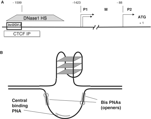
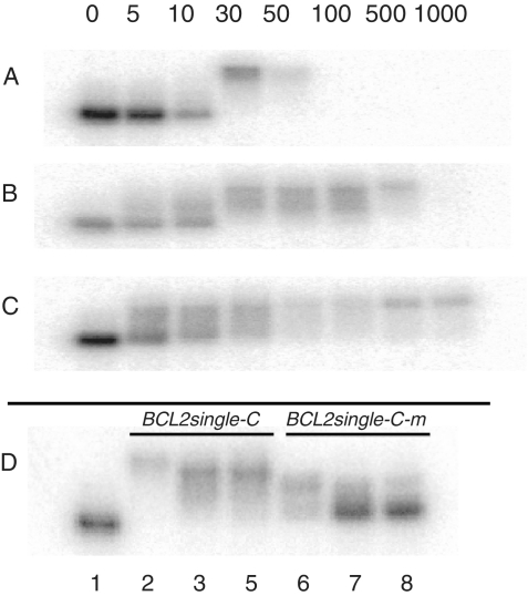
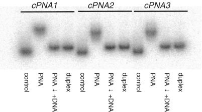
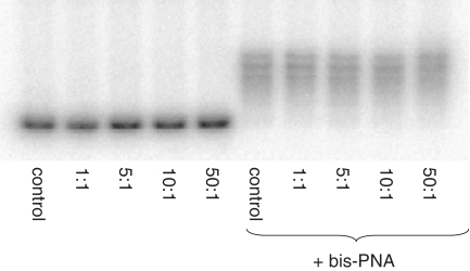
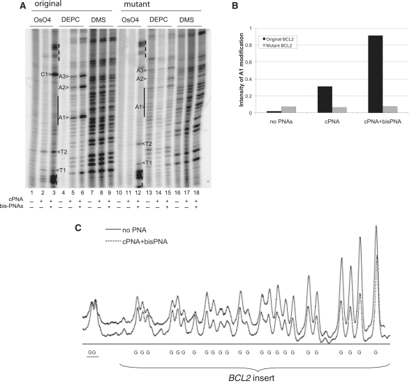
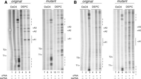
Guanine-rich sequences are highly abundant in the human genome, especially in regulatory regions. Because guanine-rich sequences have the unique ability to form G-quadruplexes, these structures may play a role in the regulation of gene transcription. In previous studies, we demonstrated that formation of G-quadruplexes could be induced with peptide nucleic acids (PNAs). PNAs designed to bind the C-rich strand upstream of the human BCL2 gene promoted quadruplex formation in the complementary G-rich strand. However, the question whether G-quadruplex formation was essential for PNA invasion remained unanswered. In this study, we compared PNA invasion in the native and mutant, i.e. not forming G-quadruplex, BCL2 sequences and showed that G-quadruplex is required for effective PNA invasion into duplex DNA. This finding provides strong evidence for not only sequence-specific, but also quadruplex specific, gene targeting with PNA probes. In addition, we examined DNA-duplex invasion potential of PNAs of various charges. Using the gel shift assay, chemical probing and dimethyl sulfate (DMS) protection studies, we determined that uncharged zwitterionic PNA has the highest binding specificity while preserving efficient duplex invasion.


Figures

Figure 1.
Figure 1.
(A) Promoter structure of the human BCL2 gene; shown bcl2G4-2 insert is a 51-mer sequence of guanine-rich strand. DNase 1 hypersensitive site (ENCODE: UW Digital Genomic Footprinting K562 cells) and CTCF binding site (ENCODE: UW Histone ChIP Raw signal CTCF in GM12878 cells) are shown as boxes. (B) Proposed design for PNA binding to G-rich DNA region.
Figure 2.
Figure 2.
Gel shift studies of single stranded DNA oligonucleotides bound with PNAs. 5′-32P labeled BCL2single-C was incubated with cPNA1 (A), cPNA2 (B) and cPNA3 (C) in TE buffer with 20 mM KCl (pH 7.4) at 37°C overnight at varying molar ratios; numbers on the top indicate PNA concentration; DNA concentration was 5 nM. Samples were run in 20% native polyacrylamide TBE gels at room temperature. (D) Binding specificity studies. 5′-32P labeled BCL2single-C and BCL2single-C-m were incubated with cPNA1 (2, 6), cPNA2 (3, 7) and cPNA3 (4, 8) in TE buffer with 20 mM KCl (pH 7.4) at 37°C overnight at 30 nM PNA and 5 nM DNA concentration; BCL2single-C without PNAs was used as a control (1).
Figure 3.
Figure 3.
Concurrent binding studies. cPNA1, cPNA2 and cPNA3 were bound to 5′-32P labeled BCL2single-C at 10:1 PNA/DNA molar ratio (lanes marked ‘PNA’). BCL2single-G was added to PNA–DNA complexes at BCL2single-G/BCL2single-C molar ratio 10:1 and incubated at 37°C overnight (lanes marked ‘PNA ↓ +DNA’). Lanes marked ‘control’ contained BCL2single-C and lanes marked ‘duplex’ contained BCL2duplex oligonucleotides. Samples were run in 20% native polyacrylamide TBE gels at room temperature.
Figure 4.
Figure 4.
Gel shift studies of plasmid DNA bound with PNAs. After incubation with PNAs (TE plus 20 mM KCl, pH 7.4, 37°C, overnight) at varying PNA molar ratios, with (right) or without (left) bis-PNA, plasmids were cut with SpeI and Pst1 restriction enzymes followed by 3′-32P labeling with [α-32P]-dCTP (Perkin Elmer) by Klenow fragment of DNA polymerase I. After purification on G-50 microcolumns samples were analyzed in native 6% polyacrylamide gel electrophoresis.
Figure 5.
Figure 5.
cPNA2 invasion studies. (A) Chemical probing of plasmid with BCL2 insert incubated with cPNA2 only and with both cPNA2 and bis-PNA. Incubation of plasmids with PNAs was performed in 20 mM KCl (pH 7.4) at 37°C overnight with molar ratio 100:1 PNA to DNA. Control sample was incubated only in TE buffer. Samples were chemically modified to a total volume of 50 μl with 2.5 mM OsO4 plus 2.5 mM 2.2′-dipyridyl disulfide for 5 min at room temperature, 2 μl DEPC for 5 min at room temperature, 2 μl 10% DMS in ethanol for 15 min at 15°C. Plasmids with two BCL2 insert were tested: with original quadruplex forming BCL2 sequence and with mutant BCL2 sequence not forming quadruplex. G-rich strand was analyzed. (B) A1 adenine modification. Intensity of the band corresponding to A1 adenine was normalized on the intensity of whole lane with background subtraction and plotted. (C) DMS modification profiles for the plasmid with original BCL2 insert incubated alone or in the presence of cPNA2 and bis-PNA. The two guanines outside of the G-quadruplex-forming region are underlined.
Figure 6.
Figure 6.
PNA invasion studies. Chemical probing of plasmid with BCL2 insert with PNAs (AcPNA1; BcPNA3). Incubation of plasmids with PNAs was performed in TE plus 20 mM KCl (pH 7.4) at 37°C overnight with molar ratio 100:1 PNA to DNA. Experimental samples were incubated with central PNAs only and with both central PNA and bis-PNA. Plasmids with two BCL2 insert were tested: with original quadruplex forming BCL2 sequence and with mutant BCL2 sequence not forming quadruplex. G-rich strand was analyzed.

References

    1. Qin Y, Rezler EM, Gokhale V, Sun D, Hurley LH. Characterization of the G-quadruplexes in the duplex nuclease hypersensitive element of the PDGF-A promoter and modulation of PDGF-A promoter activity by TMPyP4. Nucleic Acids Res. 2007;35:7698–7713. - PMC - PubMed
    1. Qin Y, Hurley LH. Structures, folding patterns, and functions of intramolecular DNA G-quadruplexes found in eukaryotic promoter regions. Biochimie. 2008;90:1149–1171. - PMC - PubMed
    1. Sun D, Guo K, Rusche JJ, Hurley LH. Facilitation of a structural transition in the polypurine/polypyrimidine tract within the proximal promoter region of the human VEGF gene by the presence of potassium and G-quadruplex-interactive agents. Nucleic Acids Res. 2005;33:6070–6080. - PMC - PubMed
    1. Guo K, Gokhale V, Hurley LH, Sun D. Intramolecularly folded G-quadruplex and i-motif structures in the proximal promoter of the vascular endothelial growth factor gene. Nucleic Acids Res. 2008;36:4598–4608. - PMC - PubMed
    1. Siddiqui-Jain A, Grand CL, Bearss DJ, Hurley LH. Direct evidence for a G-quadruplex in a promoter region and its targeting with a small molecule to repress c-MYC transcription. Proc. Natl Acad. Sci. USA. 2002;99:11593–11598. - PMC - PubMed

Publication types