. 2011 Jul;85(14):7465-71.
doi: 10.1128/JVI.00088-11. Epub 2011 May 18.

Viral interleukin-10 expressed by human cytomegalovirus during the latent phase of infection modulates latently infected myeloid cell differentiation

Affiliations

Viral interleukin-10 expressed by human cytomegalovirus during the latent phase of infection modulates latently infected myeloid cell differentiation

Selmir Avdic et al. J Virol. 2011 Jul.

Abstract

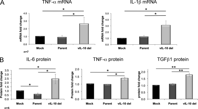
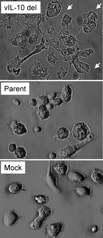
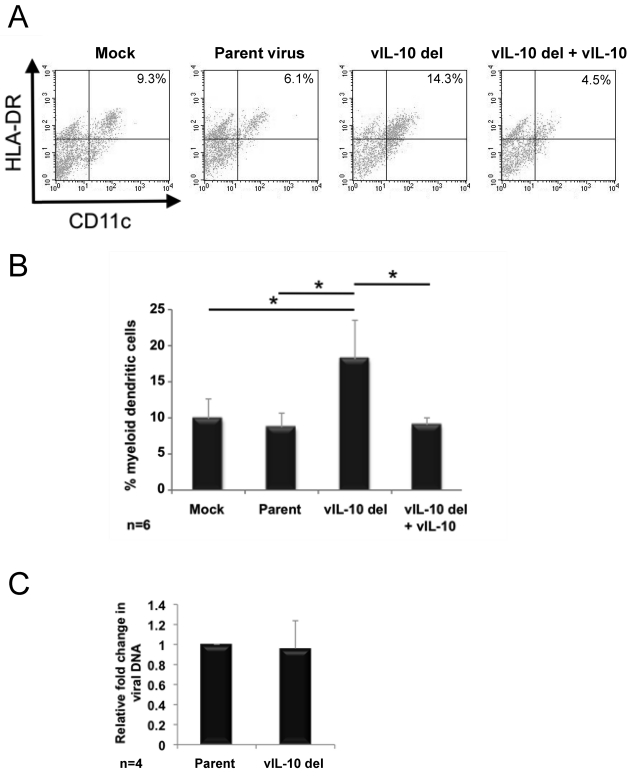
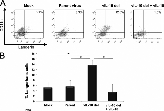
The human cytomegalovirus UL111A gene is expressed during latent and productive infections, and it codes for homologs of interleukin-10 (IL-10). We examined whether viral IL-10 expressed during latency altered differentiation of latently infected myeloid progenitors. In comparison to infection with parental virus or mock infection, latent infection with a virus in which the gene encoding viral IL-10 has been deleted upregulated cytokines associated with dendritic cell (DC) formation and increased the proportion of myeloid DCs. These data demonstrate that viral IL-10 restricts the ability of latently infected myeloid progenitors to differentiate into DCs and identifies an immunomodulatory role for viral IL-10 which may limit the host's ability to clear latent virus.


Figures

Fig. 1.
Fig. 1.
Upregulation of proinflammatory cytokines during latent infection with a viral IL-10 deletion virus. CD34+ myeloid progenitor cells were mock infected or latently infected with either parental virus (Parent) or a viral IL-10 deletion virus (vIL-10 del). (A) Fold mRNA change (relative to mock-infected cells) measured by qRT-PCR of TNF-α (primers TNF-α-F [F stands for forward] [5′-CCGTCTCCTACCAGACCAAG-3′] and TNF-α-R [R stands for reverse] [5′-CTGAGTCGGTCACCCTTCTC-3′]) and IL-1β (primers IL-1β-F [5′-GCTGAGGAAGATGCTGGTTC-3′] and IL-1β-R [5′-GTGATCGTACAGGTGCATCG-3′]) following normalization to the housekeeping gene GAPDH (primers GAPDH-F [5′-TCACCAGGGCTGCTTTTAAC-3′] and GAPDH-R [5′-GACAAGCTTCCCGTTCTCAG-3′]). (B) Fold change (relative to mock-infected cells) of secreted IL-6, TNF-α, and TGFβ1 proteins measured by ELISAs. The number of independent biological replicate experiments (n) is shown. Error bars indicate the standard errors of the means. Significant differences between values for the samples were determined by a one-tailed, paired Student's t test and are denoted by horizontal lines and asterisks as follows: *, P value of <0.05; **, P value of <0.01.
Fig. 2.
Fig. 2.
Cell morphology changes during latent infection with a viral IL-10 deletion virus. Light microscopy views of myeloid progenitor cells on day 8 after latent infection with a viral IL-10 deletion virus (vIL-10 del) or parental virus (Parent) or mock infection (Mock). The small white arrows highlight fine spiky projections on clumping cells in cultures infected with vIL-10 del virus.
Fig. 3.
Fig. 3.
Increased generation of myeloid dendritic cells from myeloid progenitor cells latently infected with a viral IL-10 deletion virus. CD34+ myeloid progenitor cells were mock infected or latently infected with either parental virus (Parent) or a viral IL-10 deletion virus (vIL-10 del). Cultures of cells infected with vIL-10 del virus supplemented with recombinant viral IL-10 proteins were also generated (vIL-10 del + vIL-10). (A) Flow cytometry scatter plots of Lin cells expressing HLA-DR and CD11c to identify myeloid DCs. (B) Graph depicting the percentage of myeloid DCs. (C) Graph depicting the fold change in viral genome load in cells infected with the viral IL-10 deletion virus relative to parental virus-infected counterparts. The number of independent biological replicate experiments (n) is shown. Error bars indicate the standard errors of the means. Significant differences (P value of <0.05) between the values for samples were determined by a one-tailed, paired Student's t test and are denoted by an asterisk.
Fig. 4.
Fig. 4.
Increased generation of Langerhans cells from myeloid progenitor cells latently infected with a viral IL-10 deletion virus. CD34+ myeloid progenitor cells were mock infected or latently infected with either parental virus (Parent) or a viral IL-10 deletion virus (vIL-10 del). Cultures of vIL-10 del virus-infected cells supplemented with recombinant viral IL-10 proteins were also generated (vIL-10 del + vIL-10). (A) Flow cytometry scatter plots of cells expressing CD11c and langerin to identify Langerhans cells. (B) Graph depicting the percentage of Langerhans cells. The number of independent biological replicate experiments (n) is shown. Error bars indicate the standard errors of the means. Significant differences (P value of <0.05) between the values for samples were determined by a one-tailed, paired Student's t test and are denoted by an asterisk.

References

    1. Anton D., Dabadghao S., Palucka K., Holm G., Yi Q. 1998. Generation of dendritic cells from peripheral blood adherent cells in medium with human serum. Scand. J. Immunol. 47:116–121 - PubMed
    1. Bego M., Maciejewski J., Khaiboullina S., Pari G., St. Jeor S. 2005. Characterization of an antisense transcript spanning the UL81-82 locus of human cytomegalovirus. J. Virol. 79:11022–11034 - PMC - PubMed
    1. Burns S., et al. 2004. Maturation of DC is associated with changes in motile characteristics and adherence. Cell Motil. Cytoskeleton 57:118–132 - PubMed
    1. Caux C., et al. 1999. Respective involvement of TGF-beta and IL-4 in the development of Langerhans cells and non-Langerhans dendritic cells from CD34+ progenitors. J. Leukoc. Biol. 66:781–791 - PubMed
    1. Chang W. L., Barry P. A., Szubin R., Wang D., Baumgarth N. 2009. Human cytomegalovirus suppresses type I interferon secretion by plasmacytoid dendritic cells through its interleukin 10 homolog. Virology 390:330–337 - PMC - PubMed

Publication types

LinkOut - more resources