Comparative Study
. 2011 May 18;31(20):7264-74.
doi: 10.1523/JNEUROSCI.6194-10.2011.

Functional alterations to the nigrostriatal system in mice lacking all three members of the synuclein family

Affiliations
Comparative Study

Functional alterations to the nigrostriatal system in mice lacking all three members of the synuclein family

Sabina Anwar et al. J Neurosci. .

Abstract

The synucleins (α, β, and γ) are highly homologous proteins thought to play a role in regulating neurotransmission and are found abundantly in presynaptic terminals. To overcome functional overlap between synuclein proteins and to understand their role in presynaptic signaling from mesostriatal dopaminergic neurons, we produced mice lacking all three members of the synuclein family. The effect on the mesostriatal system was assessed in adult (4- to 14-month-old) animals using a combination of behavioral, biochemical, histological, and electrochemical techniques. Adult triple-synuclein-null (TKO) mice displayed no overt phenotype and no change in the number of midbrain dopaminergic neurons. TKO mice were hyperactive in novel environments and exhibited elevated evoked release of dopamine in the striatum detected with fast-scan cyclic voltammetry. Elevated dopamine release was specific to the dorsal not ventral striatum and was accompanied by a decrease of dopamine tissue content. We confirmed a normal synaptic ultrastructure and a normal abundance of SNARE (soluble N-ethylmaleimide-sensitive factor attachment protein receptor) protein complexes in the dorsal striatum. Treatment of TKO animals with drugs affecting dopamine metabolism revealed normal rate of synthesis, enhanced turnover, and reduced presynaptic striatal dopamine stores. Our data uniquely reveal the importance of the synuclein proteins in regulating neurotransmitter release from specific populations of midbrain dopamine neurons through mechanisms that differ from those reported in other neurons. The finding that the complete loss of synucleins leads to changes in dopamine handling by presynaptic terminals specifically in those regions preferentially vulnerable in Parkinson's disease may ultimately inform on the selectivity of the disease process.


Figures

Figure 1.
Figure 1.
Triple-synuclein-null mutant mice possess a normal complement of dopaminergic neurons in the SNpc and VTA, normal rate of in vivo TH activity, but reduced level of dopamine and its metabolites in the striatum. A, The bar charts show means ± SEM of total number of TH-positive neurons in SNpc and VTA of 4-month-old wild-type (WT) (n = 18) and TKO (α−/−β−/−γ−/−) (n = 28) mice. Neurons were stereologically counted separately in the left and right structures. B, Striatal concentrations (picomoles per milligram of protein) of dopamine (DA) and its metabolites, DOPAC and HVA, were normalized to the mean value for wild-type animals (100%). Means ± SEM of data obtained from 15 wild-type (WT) and 14 TKO (α−/−β−/− γ−/−) mouse samples are shown (**p < 0.01; Kolmogorov–Smirnov test). For metabolite/dopamine ratios, see table (see Notes). C, The rate of l-DOPA accumulation in the striatum of wild-type (WT) (n = 5) and TKO mice (α−/−β−/−γ−/−) (n = 5) after intraperitoneal injection of 100 mg/kg AADC inhibitor NSD-1015.
Figure 2.
Figure 2.
Normal morphology and expression of synaptic markers in the striatum of triple-synuclein-null mutant mice. A, TH and DAT expression in the striatum of 4-month-old mice. Representative microphotographs of coronal sections of wild-type (WT) and TKO (α−/−β−/−γ−/−) mouse brains at the bregma 0.38 mm level. Scale bar, 1 mm (for all images). B, High-magnification confocal images of striatal sections immunofluorescently stained with antibodies against synapsin IIa. Scale bar, 2 μm (for both images). C, Western blot analysis of proteins in the striatum of wild-type and mutant mice. Representative Western blots show analysis of striatal samples from two mice for each genotype.
Figure 3.
Figure 3.
Performance of wild-type and triple-synuclein-null mutant mice in balance/coordination and exploratory behavior tests and their activity in novel nonanxiogenic environment. The bar charts show means ± SEM of experimental values obtained by testing wild-type (WT) and TKO (α−/−β−/−γ−/−) mice. For all panels, the number of animals tested and, where appropriate, a statistically significant difference are shown (*p < 0.05; **p < 0.01; Kolmogorov–Smirnov test). A, The latency to fall from the inverted grid of male 2-year-old wild-type and mutant mice of various ages. The number of animals that successfully completed the task at least once from three attempts is shown for each experimental group (in brackets). The best result for each mouse was used for calculating the group mean. B, The latency to fall from the accelerating rotarod of 4-month-old male mice. C, Percentage of trials in which 4-month-old female mice alternated in a T-maze. The difference between groups reached significance (p = 0.037) only when less robust Mann–Whitney test but not Kolmogorov–Smirnov test was used. The number of squares 4-month-old female mice entered (D) and the number of rears (E) during 3 min testing in nonanxiogenic open field. The number of nose pokes into peripheral (F) and central (G) holes during 3 min assessing of 4-month-old female mice in the holeboard test. H, I, The locomotor activity (number of infrared beam breaks) of 4-month-old male wild-type (white diamonds; n = 13) and mutant (gray squares; n = 13) mice in a home-like cage monitored for 28 h. Total number of breaks for each 1 h interval (H) and for the first or last 4 h intervals (I), both corresponding to 10:00 A.M. to 2:00 P.M., are shown. Note that a sharp increase of animal activity during the 10th interval was triggered by switching off the room light.
Figure 4.
Figure 4.
Electrically evoked dopamine transients and regulation of dopamine signals by firing frequency in wild-type and triple-synuclein-null mutant mice measured by FCV. A, Mean profiles of [DA]o versus time (mean ± SEM) after a single pulse (0.2 μs; arrow) in the dorsal striatum (CPu). In CPu, peak [DA]o transiently evoked by single pulses is greater in triple-synuclein-null mutant (α−/−β−/−γ−/−) than wild-type (WT) mice [***p < 0.001, one-way ANOVA; WT, n = 81; α−/−β−/−γ−/−, n = 86 (5 animals for each genotype)]. B, Histogram of peak [DA]o of evoked dopamine transients in the CPu in wild-type versus triple-synuclein-null mutant mice for individual sampling sites. C, Mean peak [DA]o ± SEM versus frequency during five-pulse trains (1–100 Hz) in the CPu in both genotypes (p > 0.05, two-way ANOVA; 3 animals per genotype,). D, Mean profiles of [DA]o versus time (mean ± SEM) after a single pulse (arrow) in the ventral striatum (NAc). There is no significant difference between genotype in peak evoked [DA]o [p > 0.05, one-way ANOVA; n = 26 for both genotypes (4 animals per genotype)]. E, F, Dopamine concentrations (nanograms per milligram of protein) in the CPu and NAc dissected from the striatal slices were normalized to the mean value for wild-type animals in each brain structure (100%). Means ± SEM of data obtained from 8 wild-type (WT) and 11 triple-synuclein-null mutant (α−/−β−/−γ−/−) mouse samples are shown (*p < 0.05; Kolmogorov–Smirnov test).
Figure 5.
Figure 5.
Regulation of dopamine signaling by cholinergic input, calcium, and uptake probability are similar in the CPu of wild-type and triple-synuclein-null mutant mice. A, B, Mean peak [DA]o ± SEM versus frequency during five-pulse trains (1–100 Hz) in the dorsal striatum of wild-type (A) and TKO (B) mice, with and without inhibition of nAChRs (using DHβE). Data are normalized to [DA]o under control conditions (***p < 0.001, two-way ANOVA; 3 animals per genotype). C, Response of mean peak [DA]o evoked by a single pulse to varying extracellular calcium concentrations (0.6–4.8 mm) in CPu (in the presence of DHβE) did not significantly differ between genotypes (p > 0.05, two-way ANOVA; 4 animals per genotype). Data for each genotype are normalized to mean peak [DA]o released at 4.8 mm Ca2+. Ratio of release by a four-pulse burst (100 Hz) versus a single pulse (4p:1p, right hand y-axis) as a function of calcium concentration for both genotypes (p > 0.05, two-way ANOVA; 4 animals per genotype). D, Comparison of the rates of decay of concentration-matched dopamine transients suggests that dopamine uptake rates are not significantly different between the two genotypes (p > 0.05, two-way ANOVA; n = 8 for wild-type and n = 7 for mutant mice).
Figure 6.
Figure 6.
Behavior of wild-type and triple-synuclein-null mutant mice after pharmacological challenging of dopamine neurotransmission. The locomotor activity (A–C) or climbing behavior (D) of wild-type (WT) (white diamonds) and TKO (α−/− β−/−γ−/−) (gray squares). A, Animals were injected with 4 mg/kg dAMPH after monitoring of their activity in novel environment (nonanxiogenic activity camera with infrared beams) for 30 min and returned to the same camera for an additional 90 min. Statistically significant increase in the locomotor activity of mutant mice before treatment and decrease after treatment was observed. B, A graph showing locomotor activity of triple-synuclein-null mutant mice pretreated with 50 mg/kg l-DOPA 20 min before placing in the activity camera and consequent injecting dAMPH (black circles) overlays the same graphs as in A. Statistically significant difference in the locomotor activity of wild-type and l-DOPA-pretreated mice (*) or naive and l-DOPA-pretreated mutant mice (#) is shown. C, The same protocol as described for A was used to assess locomotion of mice treated with 10 mg/kg cocaine. For all panels, statistically significant difference between two groups is shown for each 4 min interval (**,##p < 0.01; *p < 0.05; Kolmogorov–Smirnov test). D, Scoring of climbing behavior of mice after injection with 4 mg/kg APO was performed as described in Materials and Methods.
Figure 7.
Figure 7.
Electron-microscopic analyses of dopaminergic structures within the dorsal striatum in wild-type and triple-synuclein-null mutant mice. Electron micrographs of TH-immunoreactive synaptic boutons in the dorsolateral striatum of a TKO mouse. The immunoreactivity was revealed by the peroxidase method with diaminobenzidine as the chromogen (A) or the immunogold method (B). The bouton forms symmetrical synaptic contact (arrowheads) with a dendritic shaft (d). Note the nonimmunoreactive terminals forming an asymmetrical synaptic contact (white arrowheads) with a spine. Scale bars, 0.5 μm. C, Frequency distribution of the intervesicle distances in immunogold-labeled TH-positive structures (150 TH-immunoreactive profiles from 3 animals per genotype). D, Frequency distribution of the distances of vesicles from the active zone in immunogold-labeled TH-positive profiles that formed synaptic specializations (23 TH-immunoreactive synaptic boutons from 3 animals per genotype).
Figure 8.
Figure 8.
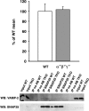
Quantification of SNARE complexes in the dorsal striatum of wild-type and triple-synuclein-null mutant mice. Coimmunoprecipitation of VAMP2/synaptobrevin with SNAP-25 was used to assess the abundance of SNARE complexes in the dorsal striatum of wild-type (WT) and TKO (α−/−β−/−γ−/−) mice. The bar chart shows amount of VAMP2/synaptobrevin in SNAP-25 immunoprecipitates normalized to the amount of precipitated SNAP-25 and expressed as percentage of mean amount WT samples (±SEM; results of 3 independent experiments; 3 WT and 3 TKO mice used in each of these experiments). A representative Western blots shows results of analysis of two independent pairs of WT and TKO mice.

References

    1. Abeliovich A, Schmitz Y, Fariñas I, Choi-Lundberg D, Ho WH, Castillo PE, Shinsky N, Verdugo JM, Armanini M, Ryan A, Hynes M, Phillips H, Sulzer D, Rosenthal A. Mice lacking alpha-synuclein display functional deficits in the nigrostriatal dopamine system. Neuron. 2000;25:239–252. - PubMed
    1. Al-Wandi A, Ninkina N, Millership S, Williamson SJ, Jones PA, Buchman VL. Absence of alpha-synuclein affects dopamine metabolism and synaptic markers in the striatum of aging mice. Neurobiol Aging. 2010;31:796–804. - PMC - PubMed
    1. Björklund A, Dunnett SB. Dopamine neuron systems in the brain: an update. Trends Neurosci. 2007;30:194–202. - PubMed
    1. Buchman VL, Hunter HJ, Pinõn LG, Thompson J, Privalova EM, Ninkina NN, Davies AM. Persyn, a member of the synuclein family, has a distinct pattern of expression in the developing nervous system. J Neurosci. 1998;18:9335–9341. - PMC - PubMed
    1. Burré J, Sharma M, Tsetsenis T, Buchman V, Etherton MR, Südhof TC. Alpha-synuclein promotes SNARE-complex assembly in vivo and in vitro. Science. 2010;329:1663–1667. - PMC - PubMed

Publication types

MeSH terms

LinkOut - more resources