Comparative Study
. 2011 May 18;31(20):7392-401.
doi: 10.1523/JNEUROSCI.6577-10.2011.

Nuclear factor erythroid 2-related factor 2 facilitates neuronal glutathione synthesis by upregulating neuronal excitatory amino acid transporter 3 expression

Affiliations
Comparative Study

Nuclear factor erythroid 2-related factor 2 facilitates neuronal glutathione synthesis by upregulating neuronal excitatory amino acid transporter 3 expression

Carole Escartin et al. J Neurosci. .

Abstract

Astrocytes support neuronal antioxidant capacity by releasing glutathione, which is cleaved to cysteine in brain extracellular space. Free cysteine is then taken up by neurons through excitatory amino acid transporter 3 [EAAT3; also termed Slc1a1 (solute carrier family 1 member 1)] to support de novo glutathione synthesis. Activation of the nuclear factor erythroid 2-related factor 2 (Nrf2)-antioxidant responsive element (ARE) pathway by oxidative stress promotes astrocyte release of glutathione, but it remains unknown how this release is coupled to neuronal glutathione synthesis. Here we evaluated transcriptional regulation of the neuronal cysteine transporter EAAT3 by the Nrf2-ARE pathway. Nrf2 activators and Nrf2 overexpression both produced EAAT3 transcriptional activation in C6 cells. A conserved ARE-related sequence was found in the EAAT3 promoter of several mammalian species. This ARE-related sequence was bound by Nrf2 in mouse neurons in vivo as observed by chromatin immunoprecipitation. Chemical activation of the Nrf2-ARE pathway in mouse brain increased both neuronal EAAT3 levels and neuronal glutathione content, and these effects were abrogated in mice genetically deficient in either Nrf2 or EAAT3. Selective overexpression of Nrf2 in brain neurons by lentiviral gene transfer was sufficient to upregulate both neuronal EAAT3 protein and glutathione content. These findings identify a mechanism whereby Nrf2 activation can coordinate astrocyte glutathione release with neuronal glutathione synthesis through transcriptional upregulation of neuronal EAAT3 expression.


Figures
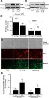
Figure 1.
Figure 1.
Activators of the Nrf2-ARE pathway increase EAAT3 expression at the transcriptional level. A, B, Cells were exposed for 48 h to SR or t-BHQ, and the nuclear accumulation of Nrf2 (A) and EAAT3 expression (B) were assessed by Western blot. C, Nrf2 and EAAT3 band densities were normalized to the corresponding PARP-1 and actin band densities, respectively, and expressed as the percentage increase over controls (CTR). n = 3–8. *p < 0.05, **p < 0.005 versus CTR. D, Immunostaining for EAAT3 and HO-1 in cells exposed to SR or t-BHQ. Scale bar, 10 μm. E, EAAT3 promoter activity was assessed using cells exposed to SR and t-BHQ and transfected with the pEAAT3-luc and pCMV-RL plasmids. n = 3–4. *p < 0.05 versus CTR.
Figure 2.
Figure 2.
Nrf2 overexpression increases EAAT3 transcription. Cells were transfected for 24 h with a plasmid encoding Nrf2, a dominant-negative Nrf2 mutant (Nrf2M), or the empty vector [control (CTR)]. Protein expression was assessed by Western blot, and band density was normalized to actin and expressed relative to CTR. n = 4–5. **p < 0.01 versus CTR; *p < 0.05 versus Nrf2.
Figure 3.
Figure 3.
Nrf2 binds to the ARE-related sequence in the EAAT3 promoter in neurons. A, The ARE-related sequence (box) is located in a conserved promoter region of the EAAT3/Slc1a1 gene in various mammalian species. The position of the first base of the ARE-related sequence (*) is given relative to the start codon for each species. The core sequence is highlighted in gray. B, Lentiviral vectors infect neurons selectively. After a double infection with lenti-LacZ and lenti-GFP, cells that are GFP and β-galactosidase positive coexpress the neuronal marker NeuN and not the astrocytic marker GFAP. Scale bar, 20 μm. C, Western blotting of brain samples from mice injected with lenti-Nrf2-HA. Antibody to the HA tag detects a band corresponding to Nrf2 only in the brains injected with lenti-Nrf2-HA. D, Chromatin from lenti-Nrf2-HA injected mice was immunoprecipitated with IgG antibody to HA or with control IgG, and the ARE-related sequence on the EAAT3 promoter was amplified by PCR using specific primers. Nrf2 binds selectively to the ARE-related sequence on the EAAT3 mouse promoter. Image is representative of three PCR runs.
Figure 4.
Figure 4.
PQT increases brain EAAT3 mRNA levels in an Nrf2-dependent manner. WT and Nrf2−/− mice received an intraperitoneal injection of 10 mg/kg PQT or saline vehicle (Veh), and the frontal cerebral cortex was collected 24 h later. AC, EAAT3 (A), HO-1 (B), and NR2B (C) mRNA levels were analyzed by qRT-PCR and normalized to GAPDH mRNA levels. n = 3–5. *p < 0.05 versus all groups.
Figure 5.
Figure 5.
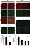
t-BHQ increases neuronal EAAT3 expression and GSH levels in an Nrf2- and EAAT3-dependent manner. Striata of WT, Nrf2−/−, and EAAT3−/− mice were injected with 1 μl of 100 mm t-BHQ or 10% ethanol vehicle (Veh) and harvested 48 h later. Photomicrographs show representative fields of the striatum near the injection sites, with EAAT3 identified by immunostaining (red) and GSH identified with C5-maleimide (green). A, In WT mice, both GSH and EAAT3 expression are upregulated by t-BHQ. B, Both basal C5-maleimide staining (C5) and the t-BHQ-mediated increase in C5-maleimide staining are suppressed in mice that also received intraperitoneal injections of BSO. C, D, t-BHQ failed to increase neuronal EAAT3 expression and GSH content in Nrf2−/− mice (C), EAAT3−/− mice (D), and in regions distant from the injection sites in WT mice (data not shown). Scale bar: (in D) AD, 40 μm. E, F, Quantification of EAAT3 and GSH staining shows that t-BHQ increases EAAT3 and GSH only in WT mice. n = 3–5. **p < 0.005, *p < 0.05, versus all groups; #p < 0.05, ##p < 0.005 versus WT Veh.
Figure 6.
Figure 6.
t-BHQ increases GSH levels in an Nrf2- and EAAT3-dependent manner. A, Striata from the mice shown in Figure 5 were also evaluated for GSH content using immunostaining for GSH-NEM adducts (green) following treatment of the sections with NEM. The increased GSH signal induced by t-BHQ was prevented by BSO and was absent at sites remote from the t-BHQ injections (data not shown). Images are representative of n = 3–5 mice in each treatment group. Scale bar, 40 μm. B, Quantified immunostaining. n = 3–5. **p < 0.01, *p < 0.05 versus Veh in each genotype.
Figure 7.
Figure 7.
Lentivirus-mediated Nrf2 overexpression in neurons increases EAAT3 expression and GSH levels. WT mice received an injection of lenti-Nrf2 in the right striatum and lenti-LacZ (control) in the left striatum. Lenti-GFP was coinjected on each side to detect infected neurons, and brains were evaluated 1 month later. A, Lenti-Nrf2 increases Nrf2 and EAAT3 mRNA levels, n = 4. *p < 0.05 versus lenti-LacZ. B, Lenti-Nrf2 increases EAAT3 expression (red) in infected neurons (green). C, Lenti-Nrf2 increases neuronal GSH content (C5 maleimide staining; red) in infected neurons. Arrows show locations of GFP-labeled neuronal perikarya in the sections injected with lenti-Nrf2. Scale bar, 40 μm. D, EAAT3 and C5 maleimide staining intensity in GFP+ (infected) neurons. n = 4. *p < 0.05.
Figure 8.
Figure 8.
Nrf2 coordinates a multicellular response to oxidative stress in the brain. Nrf2 effects on astrocytes are well described (Shih et al., 2003; Vargas and Johnson, 2009): upon stimulation by reactive oxygen species (ROS) or by Nrf2 activators such as t-BHQ and SR, Nrf2 translocates to the astrocyte nucleus (1), binds to ARE sequences, and activates the transcription of multiple genes involved in GSH metabolism. These include the cystine (Cys-Cys) transporter xCT, the GSH extruder MRP1, and the GSH degrading ecto-enzyme γ-GT (2). The net effect is increased synthesis and release of GSH by astrocytes (3). GSH is cleaved by γ-GT and aminopeptidase (AP) to generate free cysteine (Cys), the rate-limiting precursor for GSH synthesis (4). Taking advantage of a lentiviral vector that targets neurons (lenti-Nrf2, 2′), we show that Nrf2 binds to an ARE-related sequence (AREr) in the EAAT3 promoter in neurons. Activation of neuronal Nrf2 by ROS, nucleophiles (1′), or Nrf2 overexpression (2′) activates EAAT3 transcription (3′). Increased EAAT3 levels facilitate neuronal uptake of cysteine (4′), thereby facilitating GSH production in neurons (5′).

Similar articles

Cited by

References

    1. Alam J, Stewart D, Touchard C, Boinapally S, Choi AM, Cook JL. Nrf2, a Cap‘n’Collar transcription factor, regulates induction of the heme oxygenase-1 gene. J Biol Chem. 1999;274:26071–26078. - PubMed
    1. Aoyama K, Suh SW, Hamby AM, Liu J, Chan WY, Chen Y, Swanson RA. Neuronal glutathione deficiency and age-dependent neurodegeneration in the EAAC1 deficient mouse. Nat Neurosci. 2006;9:119–126. - PubMed
    1. Aoyama K, Matsumura N, Watabe M, Nakaki T. Oxidative stress on EAAC1 is involved in MPTP-induced glutathione depletion and motor dysfunction. Eur J Neurosci. 2008a;27:20–30. - PubMed
    1. Aoyama K, Watabe M, Nakaki T. Regulation of neuronal glutathione synthesis. J Pharmacol Sci. 2008b;108:227–238. - PubMed
    1. Bianchi MG, Gazzola GC, Cagnin S, Kagechika H, Bussolati O. The ATRA-dependent overexpression of the glutamate transporter EAAC1 requires RARbeta induction. Biochim Biophys Acta. 2009;1788:1861–1868. - PubMed

Publication types

LinkOut - more resources