Comparative Study
. 2011 May 18;31(20):7424-40.
doi: 10.1523/JNEUROSCI.0936-11.2011.

Deletion of the hyperpolarization-activated cyclic nucleotide-gated channel auxiliary subunit TRIP8b impairs hippocampal Ih localization and function and promotes antidepressant behavior in mice

Affiliations
Comparative Study

Deletion of the hyperpolarization-activated cyclic nucleotide-gated channel auxiliary subunit TRIP8b impairs hippocampal Ih localization and function and promotes antidepressant behavior in mice

Alan S Lewis et al. J Neurosci. .

Abstract

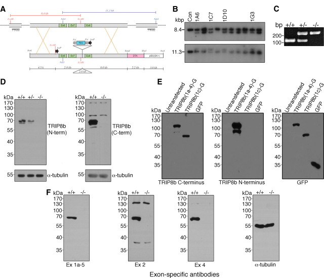
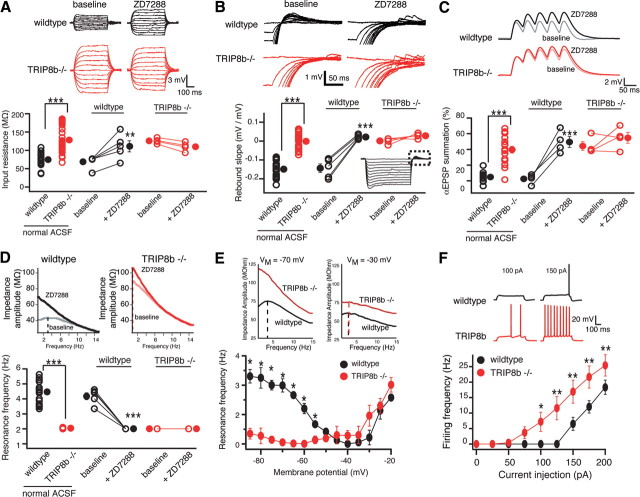
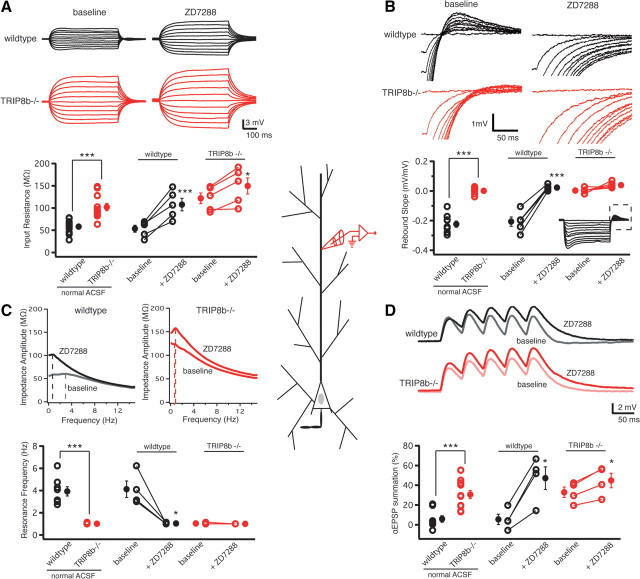
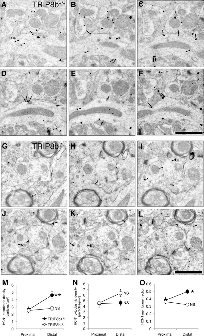
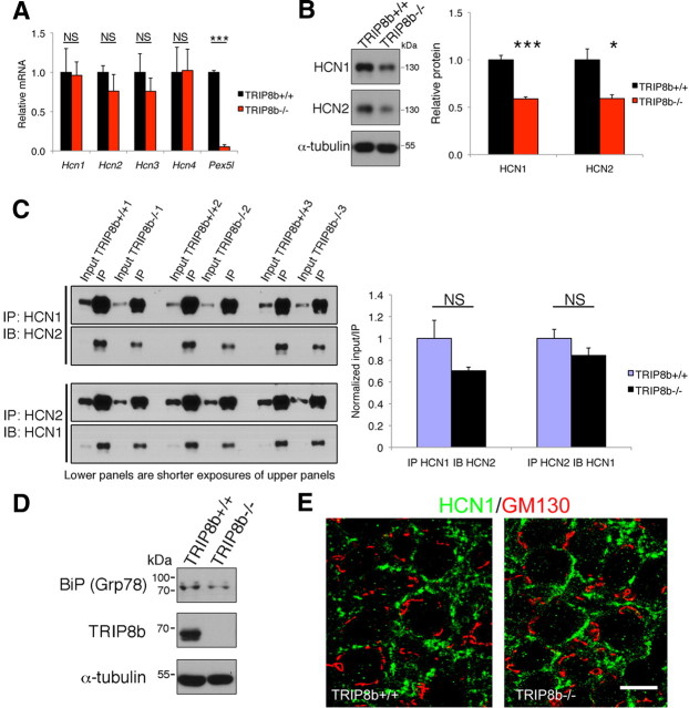
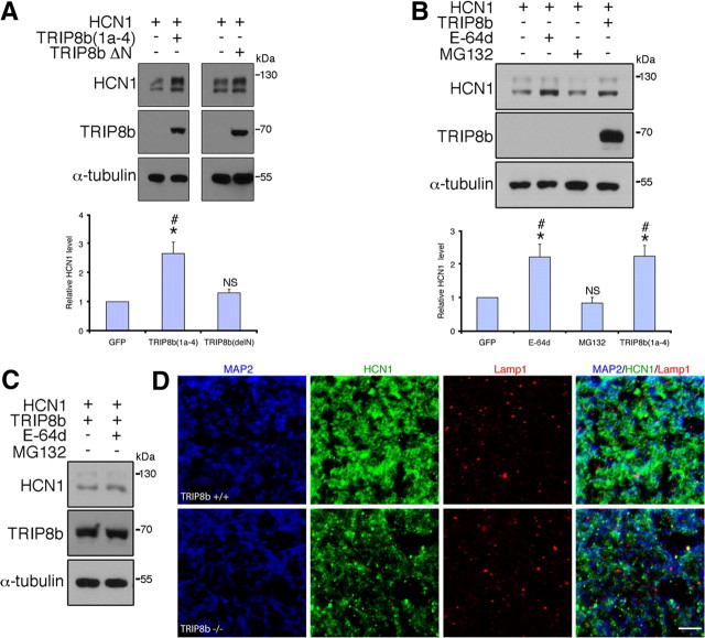
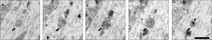
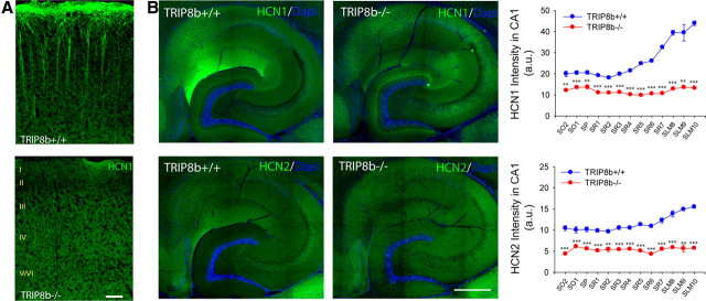
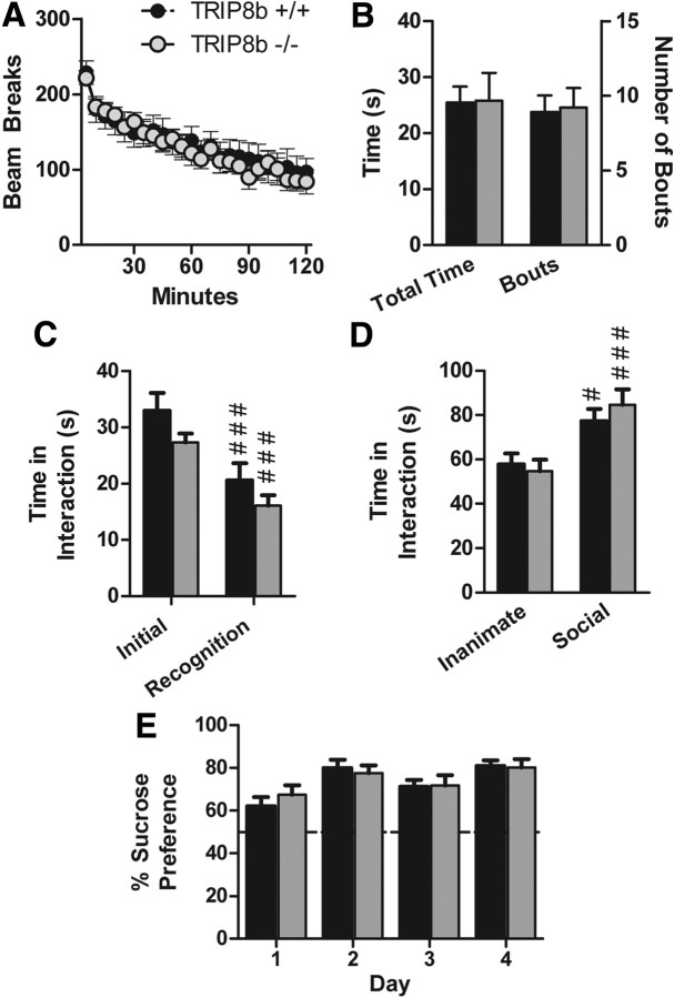
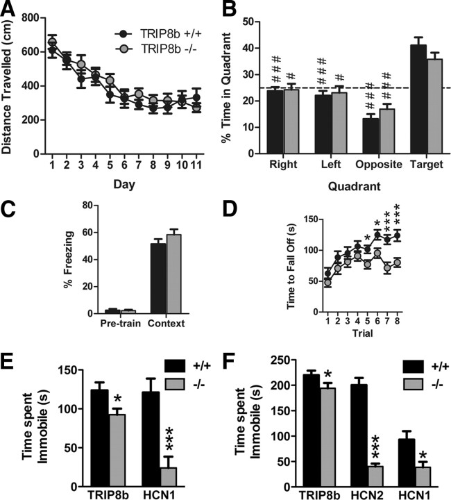
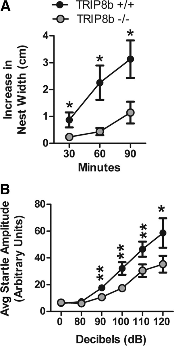
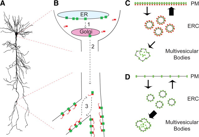
Output properties of neurons are greatly shaped by voltage-gated ion channels, whose biophysical properties and localization within axodendritic compartments serve to significantly transform the original input. The hyperpolarization-activated current, I(h), is mediated by hyperpolarization-activated cyclic nucleotide-gated (HCN) channels and plays a fundamental role in influencing neuronal excitability by regulating both membrane potential and input resistance. In neurons such as cortical and hippocampal pyramidal neurons, the subcellular localization of HCN channels plays a critical functional role, yet mechanisms controlling HCN channel trafficking are not fully understood. Because ion channel function and localization are often influenced by interacting proteins, we generated a knock-out mouse lacking the HCN channel auxiliary subunit, tetratricopeptide repeat-containing Rab8b-interacting protein (TRIP8b). Eliminating expression of TRIP8b dramatically reduced I(h) expression in hippocampal pyramidal neurons. Loss of I(h)-dependent membrane voltage properties was attributable to reduction of HCN channels on the neuronal surface, and there was a striking disruption of the normal expression pattern of HCN channels in pyramidal neuron dendrites. In heterologous cells and neurons, absence of TRIP8b increased HCN subunit targeting to and degradation by lysosomes. Mice lacking TRIP8b demonstrated motor learning deficits and enhanced resistance to multiple tasks of behavioral despair with high predictive validity for antidepressant efficacy. We observed similar resistance to behavioral despair in distinct mutant mice lacking HCN1 or HCN2. These data demonstrate that interaction with the auxiliary subunit TRIP8b is a major mechanism underlying proper expression of HCN channels and I(h) in vivo, and suggest that targeting I(h) may provide a novel approach to treatment of depression.


Figures

Figure 1.
Figure 1.
Genetic deletion of exons 6–7 of Pex5l eliminates TRIP8b protein expression in mouse brain. A, Schematic illustrating the targeted portion of Pex5l gene (top) and the targeting vector (bottom). Southern blotting probes include 500 bp fragments 5′ and 3′ to the 4.5 kb short arm and 8.0 kb long arm of the targeting vector, and detect an 8.4 kb EcoRI fragment and 11.3 kb NdeI fragment, respectively, in control (wild-type) ES cells. B, Southern blotting of EcoRI- and NdeI-digested DNA from control (lane 1) and neomycin-resistant ES cells reveals 5.8 kb EcoRI and 13.3 kb NdeI fragments reflecting proper targeting in at least four clones. C, Representative PCR genotyping products from TRIP8b+/+, TRIP8b+/−, and TRIP8b−/− DNA separated on ethidium bromide-stained 2% agarose gel. D, Western blot analysis from TRIP8b+/+, TRIP8b+/−, and TRIP8b−/− brain lysates demonstrate no TRIP8b protein or truncated TRIP8b protein using antibodies to the N terminus and C terminus. E, HEK293T cells were either untransfected or transfected with cDNAs encoding full-length TRIP8b [TRIP8b(1a-4)-GFP], TRIP8b C terminus [TRIP8b(1c)-GFP], or GFP, and lysate probed with antibodies to epitopes on the C terminus of TRIP8b (left), N terminus of TRIP8b (middle), or GFP (right). The C-terminal TRIP8b antibody properly recognized both the full-length and truncated TRIP8b isoforms, whereas the N-terminal antibody only recognized the full-length TRIP8b isoform. F, Western blot analysis confirming no expression of TRIP8b splice isoforms using splice isoform-specific antibodies (Lewis et al., 2009).
Figure 2.
Figure 2.
Deletion of TRIP8b results in functional loss of Ih in CA1 pyramidal neurons. A, Input resistance (RN) was increased in TRIP8b−/− neurons (128.23 ± 6.03 MΩ; n = 30) compared with TRIP8b+/+ (wild-type) controls (74.97 ± 3.4 MΩ; n = 23). B, Neurons from TRIP8b−/− mice had a significantly lower rebound slope (0.00 ± 0.006 mV/mV; n = 30) compared with wild-type controls (−0.14 ± 0.04 mV/mV; n = 23). The dashed box represents the portion of the traces enlarged. C, TRIP8b−/− mice displayed increased temporal summation (39.61 ± 4.31%; n = 18) compared with wild-type controls (4.75 ± 2.05%; n = 11). A–C, Inset, Average response of three traces for 500 ms current injections from −50 to +50 pA in increments of 10 pA (A), 500 ms current injections from 0 to −200 pA in increments of −20 pA (B), and five αEPSP current injections at 20 Hz (C). D, Neurons from TRIP8b−/− mice (n = 28) show no resonance (resonance frequency, fR = 1.04 ± 0.02 Hz) compared with wild-type controls (n = 23; fR = 3.47 ± 0.17 Hz). A–D, ZD7288 (20 μm) significantly altered RN (A), rebound slope (B), temporal summation (C), and fR (D), in wild-type but not TRIP8b−/− neurons. E, Plot of fR versus VM revealed that TRIP8b−/− neurons (n = 5) have a significantly lower fR at hyperpolarized voltages compared with wild-type mice (n = 5) with no significant difference at depolarized voltages. Inset, Impedance amplitude profiles for wild-type (black) and TRIP8b−/− (red) mice measured at −70 mV (left) and −30 mV (right). F, Depolarizing current injection elicited significantly more action potentials from TRIP8b−/− neurons (n = 10) compared with wild-type neurons (n = 8). In all panels, the open symbols represent individual experiments and the filled symbols represent the mean ± SEM. *p < 0.05; **p < 0.01; ***p < 0.001.
Figure 3.
Figure 3.
Functional loss of Ih because of knock-out of TRIP8b occurs in the dendrites of CA1 pyramidal neurons. A, Dendritic recordings from CA1 pyramidal neuron dendrites revealed that TRIP8b−/− mice had a significantly higher dendritic RN (102.12 ± 8.12 MΩ; n = 11) compared with TRIP8b+/+ (wild-type) controls (58.03 ± 5.05 MΩ; n = 10). B, Rebound slope measured in the dendrites of CA1 pyramidal neurons from TRIP8b−/− mice was significantly decreased (0.00 ± 0.005 mV/mV; n = 9) compared with wild-type controls (−0.22 ± 0.02 mV/mV; n = 11). The dashed box represents the portion of the traces enlarged. C, Dendritic fR in CA1 pyramidal neurons from TRIP8b−/− mice was significantly lower (1.00 Hz; n = 11) compared with wild-type controls (3.93 ± 0.41 Hz; n = 8). Inset, Impedance amplitude profiles for wild-type and TRIP8b−/− CA1 pyramidal neurons. D, CA1 pyramidal neurons from TRIP8b−/− mice showed significantly more temporal summation in the dendrites (30.56 ± 4.15%; n = 11) compared with wild-type controls (5.85 ± 3.33%; n = 8). A, B, D, Inset, Average response of three traces for 500 ms current injections from −50 to +50 pA in increments of 10 pA (A), 500 ms current injections from 0 to −200 pA in increments of −20 pA (B), and five αEPSP current injections at 20 Hz (D). A–D, ZD7288 (20 μm) significantly altered rebound slope (B) and fR (C) in wild-type but not TRIP8b−/− neurons, whereas it significantly altered RN (A) and temporal summation (D) in TRIP8b−/− as well as TRIP8b+/+ neurons. In all panels, the open symbols represent individual experiments, and the filled symbols represent the mean ± SEM. *p < 0.05; **p < 0.01; ***p < 0.001.
Figure 4.
Figure 4.
HCN1 protein density is significantly reduced on the plasma membrane of TRIP8b−/− dendrites. A–F, Serial sections through TRIP8b+/+ mouse area CA1 SLM immunostained for HCN1. HCN1 immunogold particles are found on the plasma membrane (arrows) as well as localized in the cytoplasm (arrowheads). Scale bar, 1 μm. G–L, Serial sections through TRIP8b−/− mouse area CA1 SLM immunostained for HCN1 demonstrate both significantly reduced total immunoreactivity, as well as numerous cytoplasm-localized (arrowhead) immunogold particles, although some immunogold particles are observed on the plasma membrane (arrows). Scale bar, 1 μm. Percentages of immunonegative dendrites in TRIP8b−/− mice were higher (proximal, 43%; distal, 38%) compared with TRIP8b+/+ mice (proximal, 17%; distal, 14%). M, Average HCN1 membrane particle density for TRIP8b+/+ and TRIP8b−/− proximal and distal dendrites. **p < 0.01 for TRIP8b+/+, genotype by proximal/distal interaction, F(1,151) = 8.76; multivariate analysis of covariance (MANCOVA), using dendritic diameter as covariate. N, HCN1 cytoplasmic particle density for TRIP8b+/+ and TRIP8b−/− proximal and distal dendrites. Although there was no statistically significant effect of genotype on cytoplasmic particle density, a planned comparison of TRIP8b+/+ distal and TRIP8b−/− distal cytoplasmic particle density yielded a p = 0.0586, which indicates a trend toward higher cytoplasmic immunoreactivity in the TRIP8b−/− mice. O, Quantification of fraction of HCN1 particles localized to the plasma membrane. *p < 0.05 for TRIP8b+/+, genotype by proximal/distal interaction, F(1,151) = 6.19; MANCOVA with dendritic diameter as covariate. For M–O, NS signifies p > 0.05, genotype by proximal/distal interaction, MANCOVA with dendritic diameter as covariate. For all calculations, total n = 156; TRIP8b+/+ proximal, 23; TRIP8b+/+ distal, 34; TRIP8b−/− proximal, 67; TRIP8b−/− distal, 32.
Figure 5.
Figure 5.
TRIP8b deletion reduces HCN1 and HCN2 protein levels without affecting HCN1/HCN2 heteromerization, upregulation of the unfolded protein response protein BiP (Grp78), or sequestration of HCN1 protein in Golgi apparatus. A, qPCR from hippocampal mRNA shows absence of Pex5l mRNA in TRIP8b−/− mice but no significant differences in mRNA coding for HCN channel subunits (***p < 0.001, n = 3, unpaired t test). B, Immunoblots of lysates from subdissected hippocampal area CA1 show decreased HCN1 and HCN2 subunit protein in TRIP8b−/− mice (*p < 0.05, ***p < 0.001, n = 4–6, unpaired t test). C, Immunoprecipitation (IP) of HCN1 followed by immunoblotting (IB) for HCN2 (left, top panels) and IP of HCN2 followed by IB for HCN1 (left, bottom panels) from hippocampal lysates was not significantly different in TRIP8b−/− mice compared with TRIP8b+/+ mice (n = 3 mice/genotype; p > 0.10, unpaired t test). Input is 5% of total lysate immunoprecipitated. The bottom blot shows shorter exposure of top blot, used to quantify the IP fraction compared with the input quantified from top blot. NS, Not significant. D, Immunoblotting of hippocampal lysates from TRIP8b+/+ and TRIP8b−/− mice with antibodies against BiP (Grp78), TRIP8b, and α-tubulin. E, Immunohistochemistry for HCN1 (green) and GM130 (marker of Golgi apparatus; red) in CA1 stratum pyramidale from TRIP8b+/+ and TRIP8b−/− mice demonstrates no change in colocalization, suggesting TRIP8b is not required for HCN1 transit from Golgi apparatus. Scale bar, 10 μm.
Figure 6.
Figure 6.
HCN1 is targeted to lysosomes in neurons lacking TRIP8b. A, Immunoblots from HEK293T cells cotransfected with HCN1 and TRIP8b(1a-4) or TRIP8bΔN demonstrates the TRIP8b N terminus is required for increased HCN1 protein level (*p < 0.05 vs HCN1 only, one-sample t test; #p < 0.05 vs TRIP8bΔN, unpaired t test; n = 5 sets of transfections). NS, Not significant versus HCN1 only. B, HEK293T cells expressing HCN1 and GFP were treated with E-64d (10 μm) or MG132 (10 μm) and HCN1 levels analyzed by Western blot, demonstrating treatment with E-64d enhances HCN1 protein levels similarly to TRIP8b(1a-4) (*p < 0.05 vs HCN1 control, one-sample t test; #p < 0.05 vs MG132-treatment, one-way ANOVA with Tukey's post hoc test; n = 5 sets of transfections). NS, Not significant versus control. C, HEK293T cells expressing HCN1 and TRIP8b(1a-4) were treated with vehicle or E-64d, lysed, and immunoblotted, demonstrating that lysosomal blockade does not further enhance HCN1 protein levels in TRIP8b(1a-4)-expressing cells. D, Confocal images of MAP2 (blue), HCN1 (green), and Lamp1 (red) immunostaining in SLM dendrites reveals increased colocalization of HCN1 and Lamp1 in dendrites of TRIP8b−/− mice versus controls. Scale bar, 5 μm.
Figure 7.
Figure 7.
HCN1 is localized to multivesicular bodies in distal dendrites of TRIP8b−/− mice. Representative serial sections taken from TRIP8b−/− mouse area CA1 SLM immunostained for HCN1 demonstrate immunogold localization to multivesicular bodies (MVBs) (arrows), consistent with a lysosomal degradation mechanism. HCN1 localization to MVBs was observed frequently in TRIP8b−/− SLM but rarely in TRIP8b+/+ SLM. Scale bar, 500 nm.
Figure 8.
Figure 8.
TRIP8b is required for expression of dendritic HCN channel gradients. A, Immunostaining reveals loss of HCN1 enrichment in TRIP8b−/− layer V neocortical pyramidal neuron dendrites. Scale bar, 50 μm. B, Hippocampal immunostaining for HCN1 or HCN2 (green) demonstrates loss of distal enrichment and decreased protein levels in TRIP8b−/− mice (**p < 0.01, ***p < 0.001, n = 3, unpaired t test). Regions are denoted by drawing a straight line perpendicular from the stratum pyramidale toward the apical (containing SR and SLM) or basal dendritic fields (containing SO), and dividing them into equal length sections. SO was divided into two sections, SP was divided into one section, SR was divided into seven sections, and SLM was divided into three sections. This method was adapted from the study by Shin and Chetkovich (2007). Scale bar, 400 μm.
Figure 9.
Figure 9.
TRIP8b−/− mice do not exhibit gross behavioral abnormalities. A, Locomotor activity. Data represent the number of photobeam breaks during 5 min bins. B, Grooming behavior. Data represent the total time spent grooming (left bars and y-axis) and the total number of individual grooming bouts (right bars and y-axis) during a 10 min period of observation. C, Social interaction with a juvenile (social learning). Experimental mice were placed in a novel cage with a juvenile mouse for 2 min (initial), and the test was repeated 3 d later (recognition) (###p < 0.001 compared with “initial,” planned comparisons, contrast analysis). D, Social interaction with a caged conspecific. Data represent the time spent interacting with an empty cage (inanimate target) or with a caged sex-matched conspecific (social target). #p < 0.05, ###p < 0.001 compared with the inanimate target, planned comparisons, contrast analysis. E, Sucrose preference test. Data represent the percentage of the total daily volume of liquid (sucrose or water) consumed that was consumed from the sucrose bottle (volume of sucrose solution consumed/total volume consumed). The dotted line represents chance performance. All data are from 19 littermate pairs.
Figure 10.
Figure 10.
TRIP8b−/−, HCN2ap/ap, and HCN1−/− mice exhibit reduced depression-like behavior, and TRIP8b−/− mice exhibit impaired motor learning and normal hippocampal-dependent learning and memory. A, Water maze training trials. Distance traveled to reach the hidden platform over time (n = 18 littermate pairs). B, Water maze probe trial. Percentage time spent in each quadrant of the maze (n = 18 littermate pairs; #p < 0.05, ###p < 0.001 vs target quadrant, planned comparisons, contrast analysis). C, Contextual fear conditioning. Percentage time spent freezing during initial habituation period (pretrain) and during contextual memory test (context) 24 h later (n = 19 littermate pairs). D, Rotarod. Time to fall off accelerating rotating rod (n = 19 littermate pairs; *p < 0.05, ***p < 0.001 vs TRIP8b+/+, planned comparisons, contrast analysis). E, FST on TRIP8b−/− and HCN1−/− mice along with respective controls. Total time immobile [*p < 0.05, TRIP8b−/− vs TRIP8b+/+, n = 19 littermate pairs, two-way ANOVA, main effect of genotype; ***p < 0.001, HCN1−/− vs HCN1+/+, n = 14 (HCN1−/−); n = 16 (HCN1+/+), unpaired t test]. F, TST on TRIP8b−/−, HCN1−/−, and HCN2ap/ap mice along with respective controls. Total time immobile [*p < 0.05, TRIP8b−/− vs TRIP8b+/+, n = 19 littermate pairs, two-way ANOVA, main effect of genotype; *p < 0.05, HCN1−/− vs HCN1+/+, n = 15 (HCN1−/−), n = 17 (HCN1+/+), unpaired t test; ***p < 0.001, HCN2ap/ap vs HCN2+/+, n = 7 each, unpaired t test].
Figure 11.
Figure 11.
TRIP8b−/− mice exhibit impaired nest-building behavior and decreased auditory startle response. A, Nest-building behavior. Data represent the increase in nest width, measured at 30, 60, and 90 min after placing a new nestlet in the cage (n = 19 littermate pairs; *p < 0.05 compared with TRIP8b+/+, planned comparisons, contrast analysis). The key in A also applies to B. B, Auditory startle response. Data represent the mean startle amplitude of mice when presented with auditory tones of different decibel levels (n = 19 littermate pairs; *p < 0.05, **p < 0.01 compared with TRIP8b+/+, planned comparisons, contrast analysis).
Figure 12.
Figure 12.
Schematic model of TRIP8b function in hippocampal CA1 neurons. A, Sample CA1 pyramidal neuron. B, Schematic drawing of CA1 pyramidal neuron soma (top) and distal dendrite (bottom). HCN channels (green) are expressed in endoplasmic reticulum (ER), assembled as tetramers, sorted into Golgi (1), and then trafficked into dendrites (2) without requiring TRIP8b (red). In distal dendrites, TRIP8b facilitates targeting of HCN channels to plasma membranes by unknown mechanisms (3). C, Schematic of CA1 pyramidal neuron distal dendrite showing TRIP8b (red) colocalized with HCN channels (green) and favoring trafficking from an endocytic recycling compartment (ERC) to plasma membrane (thick arrow) (PM), and limiting trafficking to multivesicular bodies/lysosomes. D, In the TRIP8b−/− mice, HCN channels are not efficiently targeted to plasma membrane resulting in increased targeting to multivesicular bodies and degradation by lysosomes (thick arrow).

References

    1. Amery L, Sano H, Mannaerts GP, Snider J, Van Looy J, Fransen M, Van Veldhoven PP. Identification of PEX5p-related novel peroxisome-targeting signal 1 (PTS1)-binding proteins in mammals. Biochem J. 2001;357:635–646. - PMC - PubMed
    1. Antonenkov VD, Van Veldhoven PP, Waelkens E, Mannaerts GP. Substrate specificities of 3-oxoacyl-CoA thiolase A and sterol carrier protein 2/3-oxoacyl-CoA thiolase purified from normal rat liver peroxisomes. Sterol carrier protein 2/3-oxoacyl-CoA thiolase is involved in the metabolism of 2-methyl-branched fatty acids and bile acid intermediates. J Biol Chem. 1997;272:26023–26031. - PubMed
    1. Baes M, Gressens P, Baumgart E, Carmeliet P, Casteels M, Fransen M, Evrard P, Fahimi D, Declercq PE, Collen D, van Veldhoven PP, Mannaerts GP. A mouse model for Zellweger syndrome. Nat Genet. 1997;17:49–57. - PubMed
    1. Barbuti A, DiFrancesco D. Control of cardiac rate by “funny” channels in health and disease. Ann N Y Acad Sci. 2008;1123:213–223. - PubMed
    1. Baruscotti M, Barbuti A, Bucchi A. The cardiac pacemaker current. J Mol Cell Cardiol. 2010;48:55–64. - PubMed

Publication types

MeSH terms