. 2011 Jun 15;186(12):6990-8.
doi: 10.4049/jimmunol.1100396. Epub 2011 May 18.

Abnormal trafficking and degradation of TLR4 underlie the elevated inflammatory response in cystic fibrosis

Affiliations

Abnormal trafficking and degradation of TLR4 underlie the elevated inflammatory response in cystic fibrosis

Emanuela M Bruscia et al. J Immunol. .

Abstract

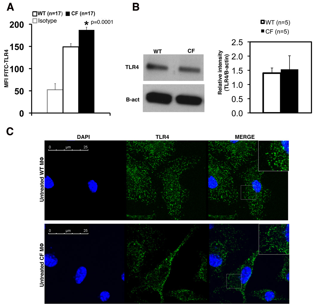
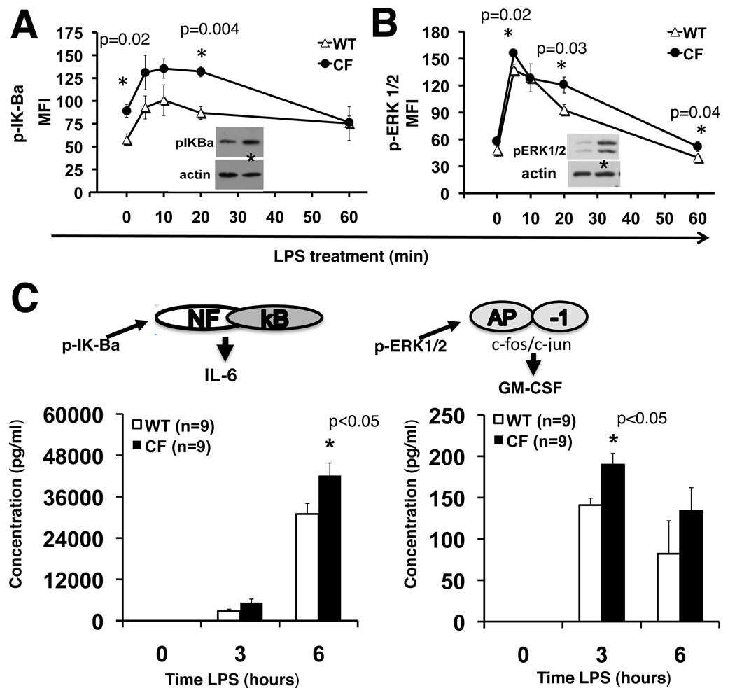
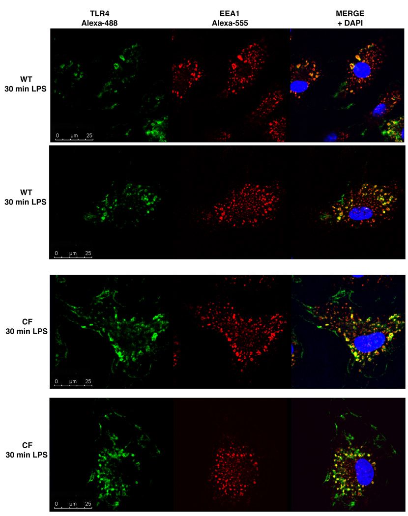
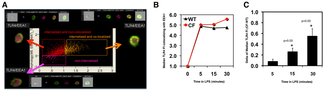
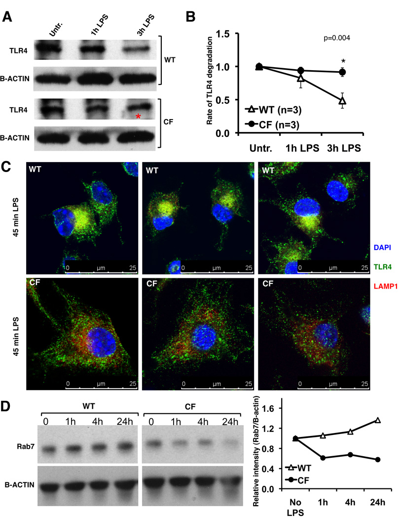
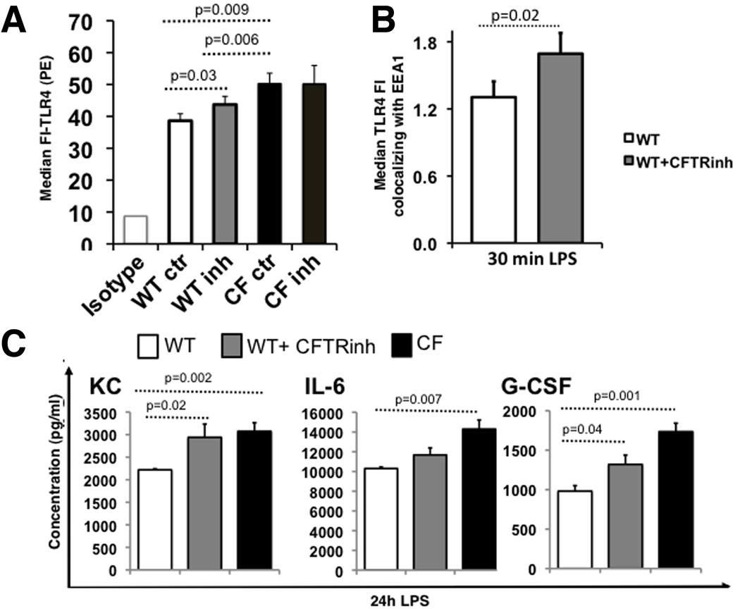
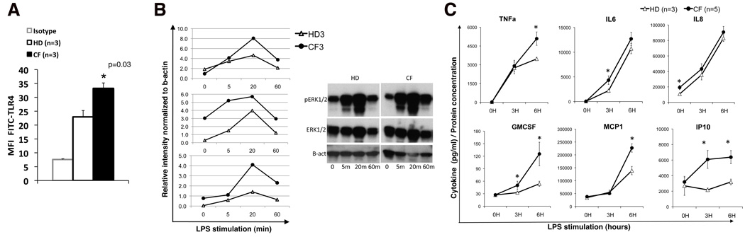
Morbidity and mortality in cystic fibrosis (CF) are due not only to abnormal epithelial cell function, but also to an abnormal immune response. We have shown previously that macrophages lacking CF transmembrane conductance regulator (CFTR), the gene mutated in CF, contribute significantly to the hyperinflammatory response observed in CF. In this study, we show that lack of functional CFTR in murine macrophages causes abnormal TLR4 subcellular localization. Upon LPS stimulation, CFTR macrophages have prolonged TLR4 retention in the early endosome and reduced translocation into the lysosomal compartment. This abnormal TLR4 trafficking leads to increased LPS-induced activation of the NF-κB, MAPK, and IFN regulatory factor-3 pathways and decreased TLR4 degradation, which affects downregulation of the proinflammatory state. In addition to primary murine cells, mononuclear cells isolated from CF patients demonstrate similar defects in response to LPS. Moreover, specific inhibition of CFTR function induces abnormal TLR4 trafficking and enhances the inflammatory response of wild-type murine cells to LPS. Thus, functional CFTR in macrophages influences TLR4 spatial and temporal localization and perturbs LPS-mediated signaling in both murine CF models and patients with CF.


Figures

Fig. 1
Fig. 1. CF macrophages have abnormal TLR4 localization compared to WT mouse macrophages
(A) Flow cytometry of plasma membrane TLR4 on untreated WT (white bars) and CF (black bars) macrophages. The isotype control is also shown. Data are representative of 17 independent experiments. (B) Western blot of total TLR4 protein from untreated WT or CF macrophages; gel is representative of 5 independent experiments; mean ± SEM relative quantification normalized to beta-actin is shown on the right. Statistical analysis was performed by two-sample t-tests. (C) Representative IF for TLR4 on untreated WT (upper panels) and CF (lower panels) macrophages; nuclei were stained with DAPI. In the merged microphotographs, the inserts represent a higher magnification of specific cell regions (gray boxes). Confocal images were taken using a 63× oil immersion lens.
Fig. 2
Fig. 2. CF macrophages have more robust and prolonged TLR4 signal transduction
(A) pIK-Balpha and (B) p-ERK1/2 assessed in untreated (0) and LPS treated (5, 10, 20 and 60 minutes) WT (open triangles) and CF (close circles) macrophages by Luminex-based phosphoprotein assay (y-axis: mean fluorescence intensity); representative western blots are also showed (20min); (C) mean ± SEM of IL-6 and GM-CSF concentration (pg/ml) in the supernatant of LPS treated WT (white bars) and CF (black bars) macrophages untreated (0) or treated with LPS for 3 and 6 hours.
Fig. 3
Fig. 3. Accumulation of activated TLR4 in the endosomal compartment of CF macrophages
After labeling plasma membrane TLR4, cells were incubated at 37°C in the presence of LPS for 30 min. Internalized TLR4 was detected by immunofluorescence for TLR4 (green), EEA-1 (red) and DAPI (blue) in 2 representative WT (upper panels) and CF (bottom panels) cells.
Fig. 4
Fig. 4. TLR4 quantification in the endosomal compartment of CF macrophages
(A) Representative Imagestream analysis, as described in the text; (B) graph from a representative experiment showing the median TLR4 fluorecence intensity (MFI) in EEA-1 positive vesicles normalized to time 0 for WT (black) and CF (red) macrophages; (C) mean ± SEM difference (Δ=CF-WT) in TLR4 MFI between WT and CF during LPS stimulation from three independent experiments. In each experiment, 5000 cells were analyzed. Statistical analysis was performed by two-sample t-tests.
Fig. 5
Fig. 5. CF macrophages have reduced TLR4 protein degradation and translocation to the lysosomal compartment
(A) Representative western blot of TLR4 from WT and CF macrophages treated with protein synthesis inhibitor and challenged with LPS. (B) Mean ± SEM relative quantification normalized to beta-actin with time zero is set at 1 for 3 independent experiments. Statistical analysis was performed by two-sample t-tests; (C) IF for TLR4 (green), LAMP-1 (red) in WT (top) and CF (bottom) macrophages treated with LPS for 45 minutes; nuclei were stained with DAPI; (D) representative western blot (left) and relative quantification normalized to b-actin (right) for Rab7 in WT and CF macrophages untreated or challenged with LPS as indicated.
Fig. 6
Fig. 6. The abnormal trafficking and signaling in CFTR−/− macrophages is CFTR-dependent
(A) TLR4 plasma membrane expression as indicated for WT, WT treated with CFTRinh172, CF and CF treated with CFTRinh172 cells: average results for 3 independent experiments (isotype control at left); (B) KC, IL-6 and G-CSF concentration (pg/ml) in the supernatant of LPS treated WT (white bars), WT treated with CFTRinh172 (gray bars) and CF (black bars) macrophages; (C) Average ± SEM MFI of TLR4 localized to the endosomal compartment after 30 minutes of LPS stimulation as assessed by Imagestream for three independent experiments (WT is white, WT treated with CFTRinh172 is light gray). Statistical analysis was performed by two-sample t-tests.
Fig. 7
Fig. 7. Human macrophages from CF patients are hyper-responsive to LPS
(A) Mean ± standard deviation of TLR4 expression in macrophages from healthy donors and patients with CF; isotype control is also shown; (B) p-ERK1/2 assessed in untreated (0) and LPS treated (5, 20 and 60 minutes) HD and CF macrophages by western blot normalized to b-actin, data are from 3 separate experiments with different CF and HD samples; a representative blot is shown (right); (C) mean ± SEM cytokine concentration in the supernatant of HD (open triangle) and CF patient (close circles) macrophages untreated (0) and treated with LPS as indicated. Statistical analysis was performed by two-sample t-tests.

References

    1. Yoshimura K, Nakamura H, Trapnell BC, Chu CS, Dalemans W, Pavirani A, Lecocq JP, Crystal RG. Expression of the cystic fibrosis transmembrane conductance regulator gene in cells of non-epithelial origin. Nucleic Acids Res. 1991;19:5417–5423. - PMC - PubMed
    1. Chmiel JF, Davis PB. State of the art: why do the lungs of patients with cystic fibrosis become infected and why can't they clear the infection? Respir Res. 2003;4:8. - PMC - PubMed
    1. Hubeau C, Puchelle E, Gaillard D. Distinct pattern of immune cell population in the lung of human fetuses with cystic fibrosis. J Allergy Clin Immunol. 2001;108:524–529. - PubMed
    1. Armstrong DS, Grimwood K, Carzino R, Carlin JB, Olinsky A, Phelan PD. Lower respiratory infection and inflammation in infants with newly diagnosed cystic fibrosis. BMJ. 1995;310:1571–1572. - PMC - PubMed
    1. Khan TZ, Wagener JS, Bost T, Martinez J, Accurso FJ, Riches DW. Early pulmonary inflammation in infants with cystic fibrosis. Am J Respir Crit Care Med. 1995;151:1075–1082. - PubMed

Publication types