. 2011 Jun 15;186(12):7144-55.
doi: 10.4049/jimmunol.1002284. Epub 2011 May 18.

Involvement of adenosine A2A receptors in engulfment-dependent apoptotic cell suppression of inflammation

Affiliations

Involvement of adenosine A2A receptors in engulfment-dependent apoptotic cell suppression of inflammation

Krisztina Köröskényi et al. J Immunol. .

Abstract

Efficient execution of apoptotic cell death followed by efficient clearance mediated by professional macrophages is a key mechanism in maintaining tissue homeostasis. Removal of apoptotic cells usually involves three central elements: 1) attraction of phagocytes via soluble "find me" signals, 2) recognition and phagocytosis via cell surface-presenting "eat me" signals, and 3) suppression or initiation of inflammatory responses depending on additional innate immune stimuli. Suppression of inflammation involves both direct inhibition of proinflammatory cytokine production and release of anti-inflammatory factors, which all contribute to the resolution of inflammation. In the current study, using wild-type and adenosine A(2A) receptor (A2AR) null mice, we investigated whether A2ARs, known to mediate anti-inflammatory signals in macrophages, participate in the apoptotic cell-mediated immunosuppression. We found that macrophages engulfing apoptotic cells release adenosine in sufficient amount to trigger A2ARs, and simultaneously increase the expression of A2ARs, as a result of possible activation of liver X receptor and peroxisome proliferators activated receptor δ. In macrophages engulfing apoptotic cells, stimulation of A2ARs suppresses the NO-dependent formation of neutrophil migration factors, such as macrophage inflammatory protein-2, using the adenylate cyclase/protein kinase A pathway. As a result, loss of A2ARs results in elevated chemoattractant secretion. This was evident as pronounced neutrophil migration upon exposure of macrophages to apoptotic cells in an in vivo peritonitis model. Altogether, our data indicate that adenosine is one of the soluble mediators released by macrophages that mediate engulfment-dependent apoptotic cell suppression of inflammation.


Figures
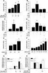
Figure 1
Figure 1. Apoptotic cell uptake leads to de novo A2A receptor synthesis in peritoneal macrophages
(A) Wild-type peritoneal macrophages were exposed for one hr. to apoptotic thymocytes alone or (B) in combination with 0.1 μg/ml cycloheximide or 10 μM actinomycin D. Thymocytes were then washed away and macrophages were further incubated (A) for the indicated time periods or for (B) 3 hrs. Cell surface expression of A2A receptor was determined by flow cytometry. (C–D) Blocking of apoptotic cell phagocytosis abolishes the increase of A2A receptor level. Phagocytosis was inhibited by pretreating macrophages with 50 mM cytochalasin D or by masking the phosphatidylserine on the apoptotic cell surface with recombinant annexin V (10 μg / 105 apoptotic cell). After one hr. co-incubation apoptotic cell were washed away and macrophages were further cultured for 2hrs (C) or 3 hrs (D). Adenosine A2A receptor level was determined by quantitative PCR (C) or by flow cytometry (D). Macrophages were treated with GW501516, a PPARδ agonist (E), or by 22 (R)OH-cholesterol, an LXR agonist (F), for 3 hrs. Cell surface expression of A2A receptor was determined by flow cytometry. A2AR gene expression level was also determined in PPARδ knock bone marrow derived (G) and LXR null peritoneal macrophages (H). Macrophages were coincubated with apoptotic thymocytes for 1 hr. Apoptotic cells were then washed away and macrophages were further incubated for 2 hrs. The gene expression levels were measured by quantitative PCR. MFI: mean fluorescence intensity. Results are expressed as mean ± S.D. of three or four independent experiments (*p<0.05).
Figure 2
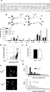
Figure 2. Adenosine A2A receptor-deficient macrophages respond to apoptotic cells with enhanced MIP-2 and KC production
(A) The map of the 40 cytokines detected on the membranes. (B) Cytokine panel of control and apoptotic cell treated wild-type and adenosine A2A receptor null peritoneal macrophages. Peritoneal macrophages were co-incubated with apoptotic thymocytes for one hr. (MPh:APO=1:10) followed by removal of apoptotic cells and addition of fresh medium. Supernatants were collected five hrs. later and cytokine levels were determined by cytokine array. Arrows highlight MIP-2, which is significantly overproduced by A2A null macrophages. (C) Cytokines, which levels were significantly different (p<0.05) in the supernatants analyzed by cytokine array. (D) Flow cytometric analysis of AnnexinV-FITC and propidium iodide stained apoptotic thymocytes. (E) Flow cytometric measurement of capacity of wild type and A2A null peritoneal macrophages to take up apoptotic thymocytes. (F–G) The number of engulfed fluorescently labeled apoptotic cells within WT or A2AR-null macrophages counted by confocal microscopy following 1h of phagocytosis. “s.b.” indicates surface bound cells. Results are expressed as mean ± S.D. of three independent experiments.
Figure 3
Figure 3. As compared to wild type controls, MIP-2 and KC production is enhanced by both A2AR null or A2AR antagonist treated wild-type macrophages while engulfing apoptotic cells
(A) Cytokine panel of control, apoptotic cell exposed and SCH442416 (A2AR-specific antagonist) treated wild-type macrophages. Peritoneal macrophages were pre-incubated with 10 nM SCH442416 for 30 min, then they were exposed to apoptotic thymocytes for one hr (MPh:APO=1:10). 5 hours after the removal of apoptotic cells supernatants were collected and cytokine levels were determined by cytokine array. Arrows highlight KC (B), MIP-2 (C), MIP-1α (D) and β (E) overproduced by SCH442416 treated macrophages. (F) MIP-2, (G) KC and (H) active TGF-β production of wild type and A2AR null macrophages engulfing apoptotic cells determined by ELISA at the same time point as above. Results are expressed as mean ± S.D. of three independent experiments (*p<0.05).
Figure 4
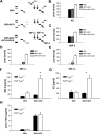
Figure 4. Increased MIP-2 production is accompanied with enhanced neutrophil migration in vivo
(A) Wild-type and A2A receptor null mice with sterile peritonitis were intraperitoneally injected with apoptotic cells (105 cells / mouse in physiological saline (apo. cell) or vehicle alone. Untreated control mice were uninjected. In some cases neutralizing MIP-2 and neutralizing KC antibodies (nMIP-2 and nKC abs) or their isotype controls were also injected. Three hrs. later peritoneal cells were collected and analyzed by flow cytometry for Gr-1-positivity. The levels of MIP-2 (B) and KC (C) in the lavage fluids were determined by ELISA at the same time point. Results are expressed as mean ± S.D. of five independent experiments (*p<0.05).
Figure 5
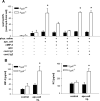
Figure 5. Transcriptionally-induced MIP-2 production by macrophages exposed to apoptotic cells is actively suppressed by the adenosine A2A receptor stimulated by adenosine in an autocrine way using the adenylate cyclase / protein kinase A signaling pathway
(A) Adenosine A2A receptor null peritoneal macrophages were exposed to apoptotic cells alone or after a 30 min. pretreatment with forskolin (10 μM), cholera toxin (100 ng / ml) or dibutyryl-cAMP (100 μM). (B) Wild-type peritoneal macrophages were exposed to apoptotic cells alone or in combination with 100 μM Rp-cAMP triethylamine or adenosine deaminase (1 U/ml). (C) Wild type or adenosine A2A receptor null peritoneal macrophages were exposed to apoptotic cells alone or after a 30 min. pretreatment with 5 μg / ml actinomycin D or 0.1 μg / ml cycloheximide. In all these experiments (A–C) apoptotic cells were washed away after one hr. and supernatants were collected after 5 hrs. to determine MIP-2 production by ELISA. (D) Quantitative RT-PCR analysis of MIP-2 mRNA expression in wild-type resting, one hr. apoptotic cell exposed peritoneal macrophages at 2 hours after the removal of the apoptotic cells or apoptotic thymocytes. Expression values are represented as mean amount of target mRNA normalized to the expression of cyclophilin. Results are expressed as mean ± S.D. of five independent experiments (*p<0.05).
Figure 6
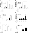
Figure 6. Apoptotic cell-induced MIP-2 production by adenosine A2A receptor null macrophages requires NO production
L-NAME, a nitric oxide synthetase inhibitor, prevents apoptotic cell-induced MIP-2 production by macrophages on both protein (A) and (B) mRNA level in a dose dependent manner. Wild type or adenosine A2A receptor null peritoneal macrophages were exposed to apoptotic cells for one hr. alone or after a 30 min. pretreatment with L-NAME in the indicated concentrations. After one hr. apoptotic cells were washed away and mRNA levels were determined 2 hrs later, while cytokine levels in the supernatant 5 hrs. later, L-NAME being constantly present. mRNA levels are expressed as fold changes as compared to the resting macrophages. (C) NO production of macrophages exposed to apoptotic cells is enhanced in the absence of A2AR. Macrophages were exposed to apoptotic cells for one hr. NO production was determined following an additional hr. (D) The NO donor sodium nitropusside enhances MIP-2 production, but only in macrophages exposed to apoptotic cells. Wild type peritoneal macrophages were exposed to the indicated concentrations of sodium nitropusside in the presence or absence of apoptotic cells. Apoptotic cells were washed away after one hr. MIP-2 mRNA levels were determined 2 hrs. later. (E) Forskolin, an adenylate cyclase activator, decreases NO production by A2AR−/− macrophages. A2AR−/− peritoneal macrophages were exposed to apoptotic cells for one hr. alone or after a 30 min. pretreatment with forskolin (10 μM). Apoptotic cells were washed away and NO production was determined following an additional hr. (F) Inhibition of protein kinase A enhances NO production of wild-type macrophages exposed to apoptotic cells. Wild-type peritoneal macrophages were exposed to apoptotic cells for one hr. alone or after a 30 min. pretreatment with 100μM Rp-cAMP triethylamine. Apoptotic cells were washed away and NO production was determined following an additional hr. Results are expressed as mean ± S.D. of five independent experiments (*p<0.05).
Figure 7
Figure 7. Loss of the adenosine A2A receptor induces changes in the mRNA expression of enzymes in arginine metabolism favoring NO production in macrophages exposed to apoptotic cells
(A) mRNA expression of arginase I and II and iNOS in resting wild type and A2AR−/− macrophages. Expression values are represented as mean amount of target mRNA normalized to the expression of cyclophilin. (B) Changes in the mRNA expression of arginase I and II and iNOS in wild type and A2AR−/− macrophages following exposure to apoptotic cells. mRNA levels are expressed as fold changes as compared to the resting macrophages. (C) Inhibition of protein kinase A in wild-type macrophages enhances, while (D) activation of adenylate cyclase in adenosine A2A receptor null macrophages decreases the mRNA levels of iNOS. The expression of arginase II (lined bars) under the same conditions is adversely affected. Results are expressed as mean ± S.D. of five independent experiments (*p<0.05).

References

    1. Savill J, Fadok V, Henson P, Haslett C. Phagocyte recognition of cells undergoing apoptosis. Immunol. Today. 1993;14:131–136. - PubMed
    1. Gregory CD, Devitt A. The macrophage and the apoptotic cell: an innate immune interaction viewed simplistically? Immunology. 2004;113:1–14. - PMC - PubMed
    1. Monoz LE, Gaipl US, Franz S, Sheriff A, Voll RE, Kalden JR, Herrmann M. SLE--a disease of clearance deficiency? Rheumatology. 2005;44:1101–1107. - PubMed
    1. Voll RE, Herrmann M, Roth EA, Stach C, Kalden JR, Girkontaite I. Immunosuppressive effects of apoptotic cells. Nature. 1997;390:350–351. - PubMed
    1. Fadok VA, Bratton DL, Konowal A, Freed PW, Wescott JY, Henson PM. Macrophages that have ingested apoptotic cells in vitro inhibit proinflammatory cytokine production through autocrine/paracrine mechanisms involving TGFβ, PGE2 and PAF. J. Clin. Invest. 1998;101:890–898. - PMC - PubMed

Publication types