Comparative Study
. 2011 Aug;106(2):801-8.
doi: 10.1152/jn.00575.2010. Epub 2011 May 18.

Nicotinic excitation of serotonergic projections from dorsal raphe to the nucleus accumbens

Affiliations
Comparative Study

Nicotinic excitation of serotonergic projections from dorsal raphe to the nucleus accumbens

Ben Chang et al. J Neurophysiol. 2011 Aug.

Abstract

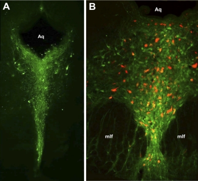
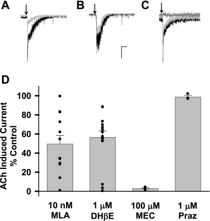
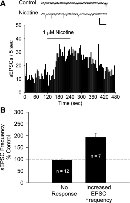
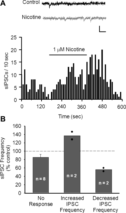
Tobacco use is a major public health problem, and although many smokers report that they want to quit, only a small percentage succeed. Side effects associated with nicotine withdrawal, including depression, anxiety, and restlessness, certainly contribute to the low success rate. The dorsal raphe nucleus (DRN) is a serotonergic center with many functions, including control of mood and emotional state. We investigated the effect of nicotine on DRN neurons that project to the nucleus accumbens (NAc), an area involved in reward-related behaviors. Using a retrograde labeling method, we found that 75% of DRN-NAc projection neurons are serotonergic. In coronal slices that include the DRN, whole cell recordings were conducted on neurons identified by fluorescent backlabeling from NAc or randomly selected within the nucleus. Nicotine increased action potential firing rates in a subset of DRN neurons. Voltage-clamp recording revealed nicotinic acetylcholine receptor (nAChR)-mediated inward currents that contribute to the nicotine-induced excitation. Nicotinic receptors also indirectly affect excitability by modulating synaptic inputs to these neurons. Nicotine enhanced excitatory glutamatergic inputs to a subset of DRN-NAc projection neurons, while inhibitory γ-aminobutyric acid (GABA)ergic inputs were modulated either positively or negatively in a subset of these neurons. The net effect of nAChR activation is enhancement of serotonergic output from DRN to the NAc, which may contribute to the effects of nicotine on mood and affect.


Figures

Fig. 1.
Fig. 1.
The dorsal raphe nucleus (DRN) projects to the nucleus accumbens (NAc). A: simplified diagram of parasagittal section illustrating DRN and ventral tegmental area (VTA) projections to NAc. PAG, periaqueductal gray. B: diagram of coronal section at the level of the NAc, bregma +1.00. C: light micrograph showing example of bilateral Fluoro-Red injection sites in NAc. Most injections occurred in both core and shell subregions. D: diagram of coronal section at the level of the PAG, bregma −7.80. E: fluorescent image showing retrograde labeling of neurons in the DRN that project to the NAc. Aq, aqueduct; mlf, medial longitudinal fasciculus. Diagrams in B and D are adapted from Paxinos and Watson (1998), Copyright Elsevier.
Fig. 2.
Fig. 2.
The majority of cells in the DRN that project to the NAc are serotonergic. A: coronal section of the PAG stained for tryptophan hydroxylase (TPH; green), demonstrating TPH-positive cells clustered in the DRN at approximately bregma −8.6. B: overlay image of retrogradely labeled DRN neurons that project to the NAc (red) and TPH-positive cells (green) at approximately bregma −7.6. In the population of backlabeled DRN-NAc projection neurons, 74.5% were colabeled for TPH (n = 1,378 backlabeled neurons from 6 rats).
Fig. 3.
Fig. 3.
Nicotine increases DRN neuronal firing rate. A: example frequency histogram from a DRN neuron that responded to bath application of 1 μM nicotine with an increase in action potential (AP) frequency. Inset: representative traces before (Control) and after perfusion with nicotine. B: mean change in firing frequency following application of 1 μM nicotine. Responsive cells were identified by comparing firing frequency before and during nicotine application (see materials and methods). Black bars represent recordings in artificial cerebrospinal fluid (aCSF), while gray bars represent recordings in aCSF + 12 μM phenylephrine. Bars represent means ± SE. C: response prevalence of the effects of 1 μM nicotine on AP frequency for cells recorded in normal aCSF (black bars) and aCSF + 12 μM phenylephrine (gray bars). B and C: numbers inside bars indicate n for each condition.
Fig. 4.
Fig. 4.
DRN neurons express functional nicotinic acetylcholine receptors (nAChRs). A: focal application of 1 mM ACh (arrows) induced rapid inward currents in 60.7% of DRN neurons tested (n = 84, black traces). In a subset of responsive neurons, pretreatment with 10 nM methyllycaconitine (MLA) inhibited ACh-induced currents (gray trace). B: in a subset of responsive neurons, pretreatment with 1 μM dihydro-β-erythroidine (DHβE) inhibited ACh-induced currents (gray trace). C: pretreatment of the slice with 100 μM mecamylamine (MEC, dark gray trace) blocked the ACh-induced current (black trace), but 1 μM prazosin (Praz, light gray trace) had no effect. D: average ACh-induced current in the presence of the antagonists tested as % of control. Bars represent means ± SE. Filled circles represent individual current amplitudes. Scale: A and B, 50 pA and 1 s; C: 200 pA, 0.5 s.
Fig. 5.
Fig. 5.
Nicotine modulates excitatory synaptic inputs to DRN neurons. A: effect of 1 μM nicotine on spontaneous excitatory postsynaptic current (sEPSC) frequency in a DRN neuron. Inset: representative traces before (Control) and after perfusion with nicotine. Scale: 50 pA, 25 ms. B: nicotine increased the sEPSC frequency in 58.3% of DRN neurons. Responsive cells were identified by comparing sEPSC frequency before and during nicotine application (see materials and methods). Bars represent means ± SE.
Fig. 6.
Fig. 6.
Nicotine induces weak modulation of inhibitory inputs to DRN neurons. A: example frequency histogram demonstrating increased spontaneous inhibitory postsynaptic current (sIPSC) frequency after application of 1 μM nicotine. Inset: representative traces before (Control) and after perfusion with nicotine. Scale: 25 pA, 25 ms. B: effect of 1 μM nicotine application on sIPSC frequency in DRN neurons. Responsive cells were identified by comparing sIPSC frequency before and during nicotine application (see materials and methods). Nicotine increased sIPSC frequency in 16.7% of cells tested and decreased sIPSC frequency in 16.7% of cells tested. Bars represent means ± SE. Filled circles represent individual observations.

References

    1. Anda RF, Williamson DF, Escobedo LG, Mast EE, Giovino GA, Remington PL. Depression and the dynamics of smoking. A national perspective. JAMA 264: 1541–1545, 1990 - PubMed
    1. Benloucif S, Keegan MJ, Galloway MP. Serotonin-facilitated dopamine release in vivo: pharmacological characterization. J Pharmacol Exp Ther 265: 373–377, 1993 - PubMed
    1. Bitner RS, Nikkel AL. Alpha-7 nicotinic receptor expression by two distinct cell types in the dorsal raphe nucleus and locus coeruleus of rat. Brain Res 938: 45–54, 2002 - PubMed
    1. Bitner RS, Nikkel AL, Curzon P, Donnelly-Roberts DL, Puttfarcken PS, Namovic M, Jacobs IC, Meyer MD, Decker MW. Reduced nicotinic receptor-mediated antinociception following in vivo antisense knock-down in rat. Brain Res 871: 66–74, 2000 - PubMed
    1. Centers for Disease Control and Prevention The Health Consequences of Smoking: a Report of the Surgeon General. Atlanta, GA: U. S. Department of Health and Human Services, 2004 - PubMed

Publication types

MeSH terms

LinkOut - more resources