. 2011 May 18;474(7352):521-5.
doi: 10.1038/nature10136.

Agonist-bound adenosine A2A receptor structures reveal common features of GPCR activation

Affiliations

Agonist-bound adenosine A2A receptor structures reveal common features of GPCR activation

Guillaume Lebon et al. Nature. .

Abstract

Adenosine receptors and β-adrenoceptors are G-protein-coupled receptors (GPCRs) that activate intracellular G proteins on binding the agonists adenosine or noradrenaline, respectively. GPCRs have similar structures consisting of seven transmembrane helices that contain well-conserved sequence motifs, indicating that they are probably activated by a common mechanism. Recent structures of β-adrenoceptors highlight residues in transmembrane region 5 that initially bind specifically to agonists rather than to antagonists, indicating that these residues have an important role in agonist-induced activation of receptors. Here we present two crystal structures of the thermostabilized human adenosine A(2A) receptor (A(2A)R-GL31) bound to its endogenous agonist adenosine and the synthetic agonist NECA. The structures represent an intermediate conformation between the inactive and active states, because they share all the features of GPCRs that are thought to be in a fully activated state, except that the cytoplasmic end of transmembrane helix 6 partially occludes the G-protein-binding site. The adenine substituent of the agonists binds in a similar fashion to the chemically related region of the inverse agonist ZM241385 (ref. 8). Both agonists contain a ribose group, not found in ZM241385, which extends deep into the ligand-binding pocket where it makes polar interactions with conserved residues in H7 (Ser 277(7.42) and His 278(7.43); superscripts refer to Ballesteros-Weinstein numbering) and non-polar interactions with residues in H3. In contrast, the inverse agonist ZM241385 does not interact with any of these residues and comparison with the agonist-bound structures indicates that ZM241385 sterically prevents the conformational change in H5 and therefore it acts as an inverse agonist. Comparison of the agonist-bound structures of A(2A)R with the agonist-bound structures of β-adrenoceptors indicates that the contraction of the ligand-binding pocket caused by the inward motion of helices 3, 5 and 7 may be a common feature in the activation of all GPCRs.


Figures

Figure 1
Figure 1
Structure of the adenosine A2A receptor bound to NECA compared to other GPCR structures. (a) The structure of NECA-bound A2AR is shown as a cartoon (yellow) aligned with the structure of A2A-T4L bound to the inverse agonist ZM241385 (blue; PDB code 3EML). NECA is shown as a space-filling model (C, green; N, blue; O, red). (b, c) Sections through the aligned receptors in (a) to highlight the differences in the intracellular face of the receptors (b) and in the ligand binding pocket (c), with the bulge in H5 shown as an inset. (d, e) Alignment of NECA-bound A2AR (yellow) with agonist-bound β2AR-Nb80 (red; PDB code 3P0G) showing the intracellular face of the receptors (d) and the ligand binding pocket (e). NECA is shown as a space-filling model in c and e. The figures were generated using CCP4mg. Analogous alignments to opsin are depicted in Supplementary Fig. 7.
Figure 2
Figure 2
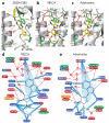
Receptor-ligand interactions compared for the adenosine receptor bound to the inverse agonist ZM241385 and the agonists NECA and adenosine. Structures of the human A2AR in cartoon representation are shown bound to the following ligands: (a) ZM241385 (PDB code 3EML); (b) NECA; (c) adenosine. (d, e) Polar and non-polar interactions involved in agonist binding to A2AR are shown for NECA (d) and adenosine (e). Amino acid residues within 3.9 Å of the ligands are depicted, with residues highlighted in blue making van der Waals contacts (blue rays) and residues highlighted in red making potential hydrogen bonds with favourable geometry (red dashed lines, as identified by HBPLUS, see Methods Online) or hydrogen bonds with unfavourable geometry (blue dashed lines, donor acceptor distance less than 3.6 Å). Where the amino acid residue differs between the human A2AR and the human A1R, A2BR and A3R, the equivalent residue is shown highlighted in orange, purple or green, respectively. Panels a-c were generated using Pymol (DeLano Scientific Ltd). Omit densities for the ligands are shown in Supplementary Fig. 6 and densities for water molecules in Supplementary Fig. 8.
Figure 3
Figure 3
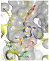
Positions of adenosine and ZM241385 in the adenosine A2A receptor ligand binding pocket. The structures of adenosine-bound A2AR-GL31 and ZM241385-bound A2A-T4L were aligned using only atoms from the protein to allow the ligand positions to be compared, with adenosine in yellow and ZM241385 in pink (N, blue; O, red). The ligands are shown in the context of the binding pocket of A2AR-GL31, with transmembrane helices of A2AR-GL31 shown in yellow and the surfaces of the receptor, including the cavity of the ligand binding pocket, shown in grey. The side chains of Val84 and Leu85 that interact with the ribose moiety of the agonist are shown in green.
Figure 4
Figure 4
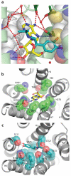
Comparison of the positions of agonists in the binding pockets of the adenosine A2A receptor and a β-adrenoceptor. (a) The structures of A2AR bound to adenosine and β1AR bound to isoprenaline (PDB code 2Y03) were aligned by superimposing equivalent atoms in the protein structure and the positions of both ligands shown as stick models with the carbon atoms in blue-green (isoprenaline) or yellow (adenosine) nitrogen in blue and oxygen in red. The A2AR structure is shown, with H5 and H7 as space-filling models (C, grey; N, blue; O, red) and the remainder of the structure as a cartoon (pale green). Some water molecules are shown as red spheres, hydrogen bonds as red dashed lines and the polar contacts as blue dashed lines. The orientation of the figure is identical to that shown in Fig. 2. (b) Structure of A2AR bound to adenosine viewed from the extracellular surface. (c) Structure of β1AR bound to isoprenaline (PDB code 2Y03) viewed from the extracellular surface. In panels b and c, equivalent side chains in the respective structures that make contacts to both isoprenaline and adenosine in their respective receptors are shown as space-filling models and they have the following Ballesteros-Weinstein numbers (amino acid side chains are shown in parentheses for the A2AR and β1AR, respectively): 3.32 (V84, D121); 3.36 (T88, V125); 5.42 (N181*, S211); 6.51 (L249, F306); 6.55 (N253, N310); 6.52 (H250*, F307); 7.39 (I274, N329); 7.43 (H278, Y333). Some residues (*) only make indirect contacts to the agonists via a water molecule.

References

    1. Potterton L, et al. Developments in the CCP4 molecular-graphics project. Acta Crystallogr D Biol Crystallogr. 2004;60:2288–2294. - PubMed
    1. Weiss HM, Grisshammer R. Purification and characterization of the human adenosine A(2a) receptor functionally expressed in Escherichia coli. Eur J Biochem. 2002;269:82–92. - PubMed
    1. Warne T, Chirnside J, Schertler GF. Expression and purification of truncated, non-glycosylated turkey beta-adrenergic receptors for crystallization. Biochim Biophys Acta. 2003;1610:133–140. - PubMed
    1. Schaffner W, Weissmann C. A rapid, sensitive, and specific method for the determination of protein in dilute solution. Anal Biochem. 1973;56:502–514. - PubMed
    1. Gorrec F, Palmer C, Lebon G, Warne T. Pi sampling: a methodical and flexible approach to macromolecular crystallization initial screening. Acta Cryst. D. 2011 In press. - PMC - PubMed

Publication types

MeSH terms