Review
. 2011 Jun;11(6):438-48.
doi: 10.1038/nrc3069. Epub 2011 May 19.

Assessing the risk of second malignancies after modern radiotherapy

Affiliations
Review

Assessing the risk of second malignancies after modern radiotherapy

Wayne D Newhauser et al. Nat Rev Cancer. 2011 Jun.

Abstract

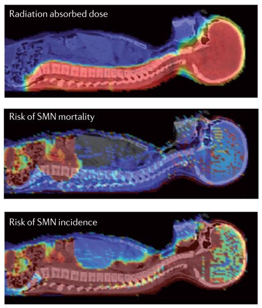
Recent advances in radiotherapy have enabled the use of different types of particles, such as protons and heavy ions, as well as refinements to the treatment of tumours with standard sources (photons). However, the risk of second cancers arising in long-term survivors continues to be a problem. The long-term risks from treatments such as particle therapy have not yet been determined and are unlikely to become apparent for many years. Therefore, there is a need to develop risk assessments based on our current knowledge of radiation-induced carcinogenesis.


Figures

Figure 1
Figure 1. Treatment planning
Examples of treatment plans for craniospinal irradiation using photon (part a) or proton (parts b and c) beams. The dose levels are shown as semi-transparent colour, superimposed on axial images of the patient from a computerized tomography scan. In the X-ray treatment (part a), the therapeutic radiation is highly localized laterally to the region of diseased tissue. Healthy tissues such as the heart and thyroid are in the beam path and receive large exit doses. For passive-scattered proton beams the therapeutic radiation (part b) is highly localized to the diseased tissues, whereas the comparatively low doses of leakage neutrons (part c) irradiate the entire body. Treatment plans for prostate radiotherapy are shown in parts d and e, with intensity-modulated radiation therapy (part d; seven coplanar fields) and a 12C ion plan (part e; two fields). Shown in greyscale is the computerized tomogram overlaid by the prescribed dose percentage in colour. The thick contour in black represents the clinical target volume, the dashed contour the gross tumour volume, and in thinner contours the rectum and the femoral head, which are tissues that are at risk from radiation. Parts a, b and c are from the M.D. Anderson Cancer Center. Parts d and e are courtesy of A. Nikoghosyan and J. Debus, Heidelberg University Hospital, Germany.
Figure 2
Figure 2. Secondary neutron dose in particle therapy
a ∣ Schematic diagram of a spinal treatment field in particle therapy. A small diameter beam of charged particles (red) enters the treatment apparatus, which spreads the beam to a clinically useful size and collimates it to spare healthy tissues. Stray neutron radiation (green) is created by proton-induced nuclear reactions in the treatment unit and in the patient. The neutron doses provide no therapeutic benefit but increase the predicted risk that a patient will develop a second cancer later in life as a result of radiation exposure. b ∣ The energy spectrum of photoneutrons produced by megavoltage X-rays and secondary neutrons produced by nuclear interactions of charged particles is complex. The figure shows recent neutron spectral measurements at the ELEKTA Linac accelerator in the Klinikum Goethe Universität of Frankfurt, Germany, operated at 25 MV, and at GSI, Darmstadt, Germany, with a 200 MeV per nucleon 12C pencil beam stopping in a water target. The energy in MeV is on the x axis in log-scale, whereas the y axis gives the number of neutrons counted per unit solid angle (in millisteradiants (msr)) and per unit dose (in Gy) to the target. Photoneutrons were measured at 10 cm or 40 cm from the target area. Secondary neutrons produced by the 12C ions were measured at two angles from the beam path (for details of the measurements see REF. 38). The yield of neutrons decreases by increasing the distance from the target or the scattering angle, but clearly X-rays produce mostly neutrons around 1 MeV, and particle therapy neutrons with energies around 100 MeV. These different spectra result in different (organ-specific) risk factors. c ∣ Neutron radiation weighting factor wR (BOX 1) is shown as a function of the neutron energy according to the latest International Commission of Radiological Protection recommendation. The most effective neutrons are considered to be those with energies around 1 MeV. Part b courtesy of C. La Tessa, GSI, Darmstadt, Germany.
Figure 3
Figure 3. Dose–response curve for carcinogenesis
a ∣ Epidemiological data in humans are mostly derived from atomic bomb (A-bomb) survivors in the dose range 0.1–2.5 Sv. At lower doses, The International Commission on Radiological Protection (ICRP) recommends a linear extrapolation (the linear-no-threshold (LNT) model). However, the non-targeted (bystander) effect or the existence of radiosensitive subpopulations may suggest that the LNT model underestimates the risk. Conversely, an adaptive response would imply that the LNT is overestimating the risk. Ongoing research has not yet clarified the importance of these mechanisms in low-dose carcinogenesis. Similarly, the extrapolation at high doses, relevant for the risk of in-field second malignant neoplasms (SMNs), is uncertain. The risk may decrease owing to cell killing (as suggested for second thyroid cancer in the Childhood Cancer Survivor Study (CCSS) study), remain linear (as it seems for central nervous system SMNs in the CCSS database) or plateau (as suggested by second lung and breast cancers in survivors of Hodgkin’s lymphoma). Cellular repopulation during and after the therapeutic radiation exposure may in fact counteract cell killing at high doses. b ∣ An example of dose–response curves for the induction of cancer in animal models by radiation of different qualities. Acute myeloid leukaemia (AML) in CBA mice exposed to γ-rays, fission neutrons or 1 GeV per nucleon 56Fe ions. The relative biological effectiveness (RBE) is about 3 for neutrons and 1 for heavy ions. However, the RBE values depend on the genetic strain, tumour type and fractionation used. The curves represent guides drawn by eye from data points in REFS 82,. Part a courtesy of E. J. Hall, Columbia University, USA.
Figure 4
Figure 4. Dose and risk distribution for second cancer
A 9-year-old girl received craniospinal irradiation for medulloblastoma using passively scattered proton beams at the M.D. Anderson Cancer Center, USA. The colour scale illustrates the difference for absorbed dose, incidence and mortality cancer risk in different organs. Radiation absorbed dose depends strongly on patient anatomy and treatment factors. Risk of second malignant neoplasm (SMN) incidence and mortality varies strongly with radiation dose, but, importantly, it also varies strongly between organs, the age of the patient at exposure and the attained age, sex and genetic profile, as well as other factors. Consequently, as this figure illustrates, dose is a poor biomarker for SMN risk. In future, novel risk visualization and analysis methods will be needed to facilitate routine risk-adapted, personalized clinical decision making.

References

    1. Smith MA, et al. Outcomes for children and adolescents with cancer: challenges for the twenty-first century. J. Clin. Oncol. 2010;28:2625–2634. - PMC - PubMed
    1. Friedman DL, et al. Subsequent neoplasms in 5-year survivors of childhood cancer: the Childhood Cancer Survivor Study. J. Natl. Cancer Inst. 2010;102:1083–1095. These are the most recent results of the CCSS, the most important epidemiological analysis of SMNs in children.

    1. Oeffinger KC, et al. for the Childhood Cancer Survivor Study Chronic health conditions in adult survivors of childhood cancer. N. Eng. J. Med. 2006;355:1572–1582. - PubMed
    1. Robison LL, et al. The Childhood Cancer Survivor Study: a National Cancer Institute-supported resource for outcome and intervention research. J. Clin. Oncol. 2009;27:2308–2318. - PMC - PubMed
    1. West C, Rosenstein BS. Establishment of a radiogenomics consortium. Radiother. Oncol. 2010;94:117–118. - PubMed

Publication types