Review
. 2011 May 19;473(7347):326-35.
doi: 10.1038/nature10147.

Heart regeneration

Affiliations
Review

Heart regeneration

Michael A Laflamme et al. Nature. .

Abstract

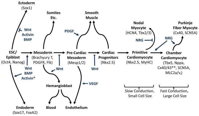
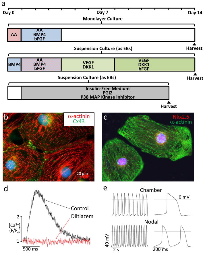
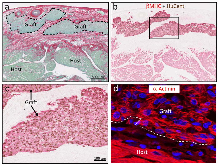
Heart failure plagues industrialized nations, killing more people than any other disease. It usually results from a deficiency of specialized cardiac muscle cells known as cardiomyocytes, and a robust therapy to regenerate lost myocardium could help millions of patients every year. Heart regeneration is well documented in amphibia and fish and in developing mammals. After birth, however, human heart regeneration becomes limited to very slow cardiomyocyte replacement. Several experimental strategies to remuscularize the injured heart using adult stem cells and pluripotent stem cells, cellular reprogramming and tissue engineering are in progress. Although many challenges remain, these interventions may eventually lead to better approaches to treat or prevent heart failure.


Figures

Figure 1
Figure 1. Cardiovascular lineages during embryonic development and embryonic stem cell differentiation
Cardiac differentiation from ESCs closely mimics cardiac development in the embryo. In either case, the specification of the cardiovascular lineages involves a transition through a sequence of increasingly restricted progenitors, proceeding from a pluripotent state to mesoderm and then to cells committed to cardiovascular fates. Growth factors that regulate fate choices are listed at branch points, and key transcription factors and surface markers for each cell state are listed under the cell types. These growth factors are useful for directing the differentiation of ESCs, while the markers are useful for purifying cells at defined developmental states. Primitive cardiomyocytes in the embryonic heart tube and nodal/pacemaker cells exhibit slow electrical propagation and a small cell size. In contrast, the eventual specification of working atrial and ventricular cardiomyocytes is accompanied by more rapid conduction, ion channel remodeling and increased cell size. While the field has made considerable progress toward the elucidation of early events during cardiogenesis, a better understanding of how pacemaker vs. chamber-specific cardiac subtypes are formed is required for clinical applications.
Figure 2
Figure 2. Guided differentiation and phenotype of cardiomyocytes from pluripotent stem cells
a, Selected protocols for the guided differentiation of human ESCs and iPSCs into cardiomyocytes using chemically-defined factors. The upper timeline shows a protocol from our group in which differentiating cells are serially pulsed with activin A (AA) and BMP4 under monolayer culture conditions. The middle timeline shows a protocol from the Keller lab that involves embryoid body (EB) formation in suspension cultures and the application of multiple signaling molecules including activin A, BMP4 The lower timeline shows a protocol from Davidson and colleagues in which EBs are continuously cultured in insulin-free medium supplemented with prostaglandin I2 and an inhibitor of p38 MAP kinase. b, Representative human ESC-derived cardiomyocyte, differentiated using the monolayer protocol, immunostained for α-actinin (red) and connexin43 (Cx43; green). c, Representative human iPSC-derived cardiomyocyte, differentiated using the monolayer protocol, immunostained for α-actinin (green) and the transcription factor Nkx2.5 (red). d, Intracellular [Ca2+] transients in a human ESC-derived cardiomyocyte before (black) and after (red) application of diltiazem, an L-type Ca2+ channel blocker. Absence of [Ca2+] transients after diltiazem treatment indicates extracellular [Ca2+] is required to initiate intracellular [Ca2+] release, just as in adult cardiomyocytes. e, Human ESC-derived cardiomyocytes show the characteristic action potential properties of either working chamber (upper) or nodal myocytes (lower), indicating early subtype specification.
Figure 3
Figure 3. Grafts of human ESC-derived cardiomyocytes in the cryoinjured guinea pig heart
Representative photomicrographs demonstrating substantial implants of human myocardium within the scar tissue. a, By picrosirius red stain, the scar appears red and viable tissue green. Scale bar=500 μm. b, The human origin of the graft myocardium was confirmed in an adjacent section by combined in situ hybridization with a human-specific pan-centromeric (HuCent, brown) probe and β-myosin heavy chain (βMHC, red) immunohistochemistry. c, Inset from panel b at higher magnification. Note the nuclear localization of the HuCent signal, confirming human origin of these cells. d, Immunostaining for α-actinin highlights the sarcomeric organization of the graft myocytes.

References

    1. Carvalho AB, de Carvalho AC. Heart regeneration: Past, present and future. World J Cardiol. 2010;2(5):107–111. - PMC - PubMed
    1. Rumyantsev PP. In: Muscle Regeneration. Mauro A, editor. Raven Press; New York: 1979. pp. 335–355.
    1. Rumyantsev PP. Growth and hyperplasia of cardiac muscle cells. Harwood Academic Publishers; 1991.
    1. Murry CE, Reinecke H, Pabon LM. Regeneration gaps: observations on stem cells and cardiac repair. J Am Coll Cardiol. 2006;47(9):1777–1785. - PubMed
    1. Whelan RS, Kaplinskiy V, Kitsis RN. Cell death in the pathogenesis of heart disease: mechanisms and significance. Annu Rev Physiol. 2010;72:19–44. - PubMed

Publication types