. 2010;2(5):587-598.

RSK2 Binding Models Delineate Key Features for Activity

Affiliations

RSK2 Binding Models Delineate Key Features for Activity

Rick Gussio et al. J Chem Pharm Res. 2010.

Abstract

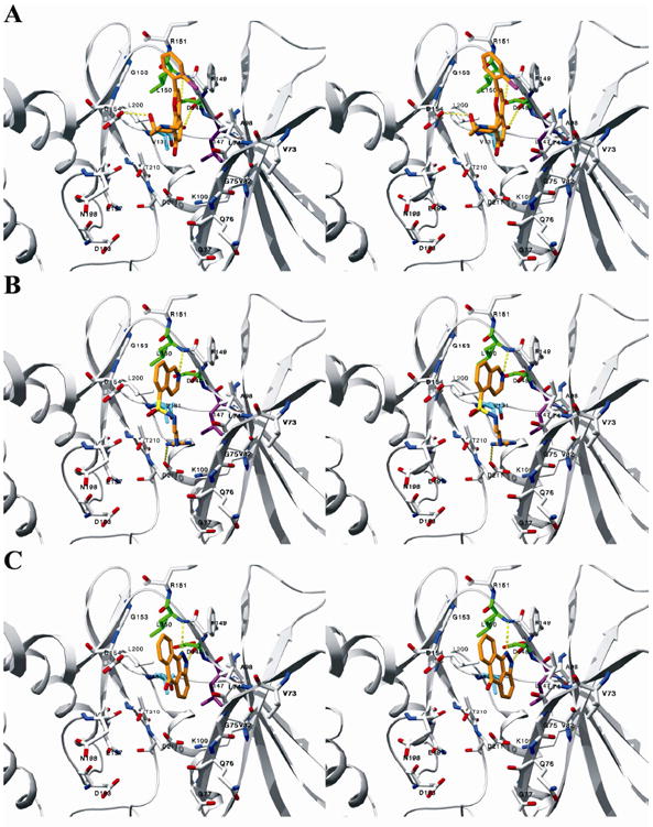
Due to its overexpression and activation in human cancer cells and tissues, an emerging molecular target in cancer therapeutics is p90 ribosomal s6 kinase 2 (RSK2). While a growing number of RSK2 inhibitors have been reported in the literature, only the crystal structure of RSK2 in complex with an AMP analogue provides a structural basis for understanding RSK2 inhibition. To remedy this, we used our fluorescence polarization assay to determine the RSK2 activity for a set of structurally diverse compounds, and followed this by modeling their binding modes in an all-atom, energy refined crystal structure of RSK2. These binding models reveal that Val131 and Leu147 are key interaction sites for potent RSK2 inhibition. This structure-based pharmacophore is an important tool for new lead discovery and refinement.


Figures

Figure 1
Figure 1
Chemical structures of RSK2 inhibitors already reported in the literature.
Figure 2
Figure 2
Chemical structures and inhibitory activities of RSK2 inhibitors used in this study.
Figure 3
Figure 3. Stereoviews of the binding models of (A) S-isomer of 2 and (B) 6
RSK2 is rendered in grey ribbon. The inhibitors and binding site residues are shown in stick. Nitrogen, oxygen and halogen atoms are colored blue, red, and purple, respectively. The carbon atoms are colored orange for the inhibitors, green for Asp148 and Leu150, which highlights the interior of the ATP-binding site, and cyan for Val131 and purple for Leu147, which demark key hydrophobic interaction sites. For clarity, hydrogen atoms are not depicted.
Figure 4
Figure 4
Stereo view of the binding models of the lower affinity inhibitors (A) 16, (B) 17, and (C) 18 rendered in the same fashion as in Figure 3.
Figure 5
Figure 5. Comparison of the RSK2 binding models of 8, 9, 12, and 19 with their counterpart X-ray structures in other serine/threonine kinases
The ligands and residues are rendered in stick. The atoms of CDK2, ROCK2, PKA, and c-JNK and their bound ligands are colored orange, while nitrogen, oxygen and carbon atoms of RSK2 and its bound ligands are colored blue, red, and green, respectively. The residue names and numberings are colored green for RSK2 and orange for CDK2, ROCK2, PK, and c-JNK. (A) Compound 8 in PKA (PDB code 1YDR). (B) Compound 9 in CDK2 (PDB code 1PF8). (C) Compound 12 in ROCK2 (PDB code 2H9V). (D) Compound 19 in c-JNK (PDB code 1UKI).

References

    1. Jones SW, Erikson E, Blenis J, Maller JL, Erikson RL. Proc Natl Acad Sci U S A. 1988;85(10):3377–81. - PMC - PubMed
    1. Alcorta DA, Crews CM, Sweet LJ, Bankston L, Jones SW, Erikson RL. Mol Cell Biol. 1989;9(9):3850–9. - PMC - PubMed
    1. Moller DE, Xia CH, Tang W, Zhu AX, Jakubowski M. Am J Physiol. 1994;266(2 Pt 1):C351–9. - PubMed
    1. Zhao Y, Bjorbaek C, Weremowicz S, Morton CC, Moller DE. Mol Cell Biol. 1995;15(8):4353–63. - PMC - PubMed
    1. Nguyen TL. Anticancer Agents Med Chem. 2008;8(7):710–6. - PubMed

LinkOut - more resources