

Identification and characterization of a neutralizing-epitope-containing spike protein fragment in turkey coronavirus
- PMID: 21594597
- PMCID: PMC7086772
- DOI: 10.1007/s00705-011-1020-1
Identification and characterization of a neutralizing-epitope-containing spike protein fragment in turkey coronavirus
Abstract
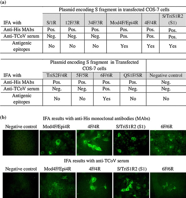
Little is known about the neutralizing epitopes in turkey coronavirus (TCoV). The spike (S) protein gene of TCoV was divided into 10 fragments to identify the antigenic region containing neutralizing epitopes. The expression and antigenicity of S fragments was confirmed by immunofluorescence antibody (IFA) assay using an anti-histidine monoclonal antibody or anti-TCoV serum. Polyclonal antibodies raised against expressed S1 (amino acid position 1 to 573 from start codon of S protein), 4F/4R (482-678), 6F/6R (830-1071), or Mod4F/Epi4R (476-520) S fragment recognized native S1 protein and TCoV in the intestines of TCoV-infected turkey embryos. Anti-TCoV serum reacted with recombinant 4F/4R, 6F/6R, and Mod4F/Epi4R in a western blot. The results of a virus neutralization assay indicated that the carboxyl terminal region of the S1 protein (Mod4F/Epi4R) or the combined carboxyl terminal S1 and amino terminal S2 protein (4F/4R) possesses the neutralizing epitopes, while the S2 fragment (6F/6R) contains antigenic epitopes but not neutralizing epitopes.
Figures


Similar articles
-
A DNA prime-protein boost vaccination strategy targeting turkey coronavirus spike protein fragment containing neutralizing epitope against infectious challenge.Vet Immunol Immunopathol. 2013 Apr 15;152(3-4):359-69. doi: 10.1016/j.vetimm.2013.01.009. Epub 2013 Feb 1. Vet Immunol Immunopathol. 2013. PMID: 23428360 Free PMC article.
-
Use of recombinant S1 spike polypeptide to develop a TCoV-specific antibody ELISA.Vet Microbiol. 2009 Sep 18;138(3-4):281-8. doi: 10.1016/j.vetmic.2009.04.010. Epub 2009 Apr 10. Vet Microbiol. 2009. PMID: 19414227 Free PMC article.
-
Application of purified recombinant antigenic spike fragments to the diagnosis of avian infectious bronchitis virus infection.Appl Microbiol Biotechnol. 2012 Jul;95(1):233-42. doi: 10.1007/s00253-012-4143-8. Epub 2012 May 25. Appl Microbiol Biotechnol. 2012. PMID: 22627759
-
Virus shedding and serum antibody responses during experimental turkey coronavirus infections in young turkey poults.Avian Pathol. 2009 Apr;38(2):181-6. doi: 10.1080/03079450902751863. Avian Pathol. 2009. PMID: 19322719
-
Recombinant nucleocapsid protein-based enzyme-linked immunosorbent assay for detection of antibody to turkey coronavirus.J Virol Methods. 2015 Jun 1;217:36-41. doi: 10.1016/j.jviromet.2015.02.024. Epub 2015 Mar 5. J Virol Methods. 2015. PMID: 25745958 Free PMC article.
Cited by
-
Unique Variants of Avian Coronaviruses from Indigenous Chickens in Kenya.Viruses. 2023 Jan 17;15(2):264. doi: 10.3390/v15020264. Viruses. 2023. PMID: 36851482 Free PMC article.
-
A DNA prime-protein boost vaccination strategy targeting turkey coronavirus spike protein fragment containing neutralizing epitope against infectious challenge.Vet Immunol Immunopathol. 2013 Apr 15;152(3-4):359-69. doi: 10.1016/j.vetimm.2013.01.009. Epub 2013 Feb 1. Vet Immunol Immunopathol. 2013. PMID: 23428360 Free PMC article.
-
Molecular Characterization of Complete Genome Sequence of an Avian Coronavirus Identified in a Backyard Chicken from Tanzania.Genes (Basel). 2023 Sep 23;14(10):1852. doi: 10.3390/genes14101852. Genes (Basel). 2023. PMID: 37895200 Free PMC article.
-
Protective Efficacy of Different Live Attenuated Infectious Bronchitis Virus Vaccination Regimes Against Challenge With IBV Variant-2 Circulating in the Middle East.Front Vet Sci. 2019 Oct 9;6:341. doi: 10.3389/fvets.2019.00341. eCollection 2019. Front Vet Sci. 2019. PMID: 31649942 Free PMC article.
-
Genotyping of turkey coronavirus field isolates from various geographic locations in the Unites States based on the spike gene.Arch Virol. 2015 Nov;160(11):2719-26. doi: 10.1007/s00705-015-2556-2. Epub 2015 Aug 8. Arch Virol. 2015. PMID: 26254026 Free PMC article.
References
-
- Guy JS. Turkey Coronavirus Enteritis. In: Saif YM, Barnes HJ, Glisson JR, Fadly AM, McDougald LR, Swayne DE, editors. Disease of poultry. 11. Ames: Iowa State University Press; 2003. pp. 300–307.
MeSH terms
Substances
LinkOut - more resources
Full Text Sources
Molecular Biology Databases
