

Case Reports
doi: 10.1007/s00586-011-1847-x.
Epub 2011 May 19.
Spinal gout tophus: a very rare cause of radiculopathy
Affiliations
- PMID: 21594750
- PMCID: PMC3369051
- DOI: 10.1007/s00586-011-1847-x
Item in Clipboard
Case Reports
Spinal gout tophus: a very rare cause of radiculopathy
Eur Spine J.
2012 Jun.
Abstract
Gout is a common metabolic disease characterized by the development of arthritis and nephropathy related to the deposition of monosodium urate crystals within the joints, periarticular tissues, skin and kidneys. Tophus formation seen around the spinal column is very rare, while occurrences of spinal gout tophus without systemic gout disease are much more unique. In our study, we report a spinal gout case that presented with right sciatica without previous history of systemic gout disease.
Figures
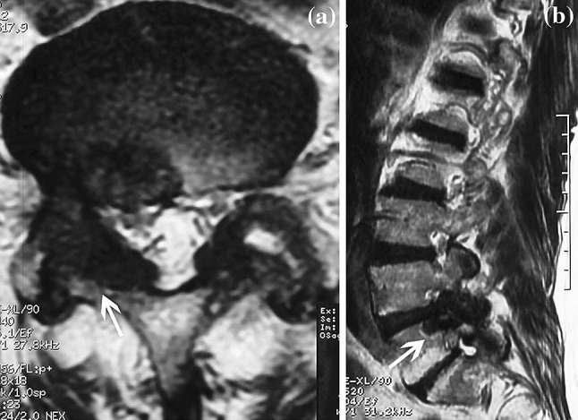
a, b T2-weighted axial and sagittal images show extradural lesions with severe dural sac and right root compression by the gout tophaceous deposits
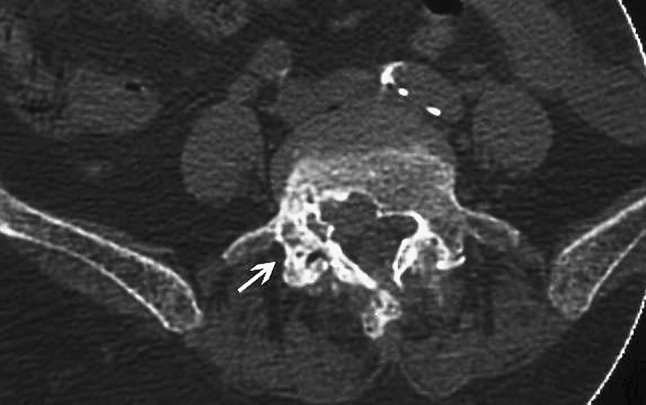
Axial CT shows bone erosions with calcifications over the right L4–5 facet joint and surrounding soft tissue masses
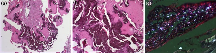
a Histologic preparation of the tissue, which shows a deposit of calcium pyrophosphate dihydrate on the soft tissue (H&E ×40). b There was a cartilaginous matrix associated with the lesion (H&E ×200). c Photomicrograph of a deposit of calcium pyrophosphate dihydrate during disease. The rhomboid-shaped crystals were examined using polarized light with red plate. The crystals are bluish white (weak positive birefringence) (×400)
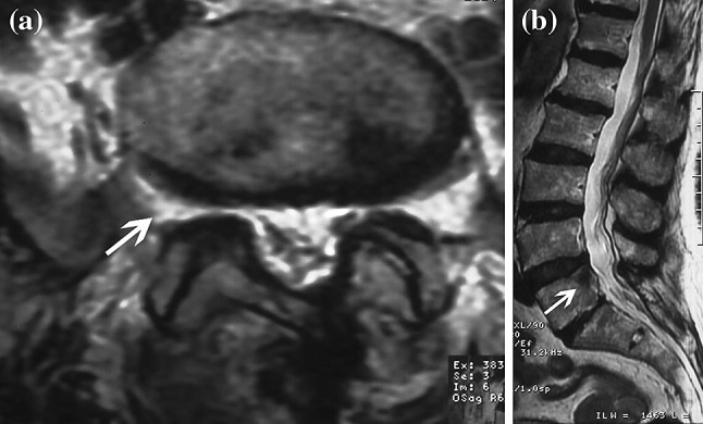
a, b Postoperative MR images
References
-
- Dhote R, Roux FX, Bachmeyer C, Tudoret L, Daumas-Duport C, Christoforov B. Extradural spinal tophaceous gout: evolution with medical treatment. Clin Exp Rheumatol. 1997;15:421–423. - PubMed
Publication types
MeSH terms
LinkOut - more resources
Full Text Sources
Medical
