

Review
doi: 10.1002/dneu.20919.
The visual system of zebrafish and its use to model human ocular diseases
Affiliations
- PMID: 21595048
- PMCID: PMC3202066
- DOI: 10.1002/dneu.20919
Item in Clipboard
Review
The visual system of zebrafish and its use to model human ocular diseases
Dev Neurobiol.
2012 Mar.
Abstract
Free swimming zebrafish larvae depend mainly on their sense of vision to evade predation and to catch prey. Hence, there is strong selective pressure on the fast maturation of visual function and indeed the visual system already supports a number of visually driven behaviors in the newly hatched larvae.The ability to exploit the genetic and embryonic accessibility of the zebrafish in combination with a behavioral assessment of visual system function has made the zebrafish a popular model to study vision and its diseases.Here, we review the anatomy, physiology, and development of the zebrafish eye as the basis to relate the contributions of the zebrafish to our understanding of human ocular diseases.
Figures
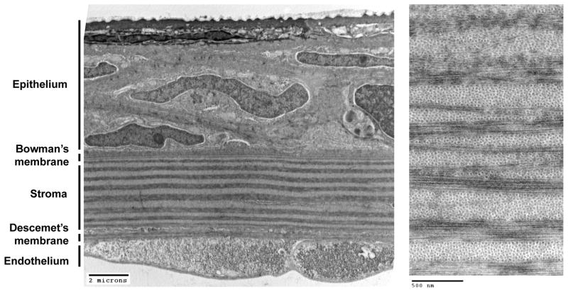
Transmission electron microscopy reveals the five principal layers of the adult zebrafish cornea (Left panel). Higher magnification of the orthogonally-arrayed collagen bundles (Right panel).
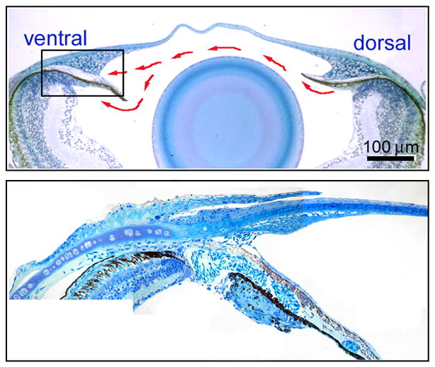
Histology showing low magnification of the adult zebrafish anterior segment (Upper panel). Red arrows show the general flow of aqueous humor. Higher magnification shows the openings of the ventral canalicular outflow pathway (Lower panel).
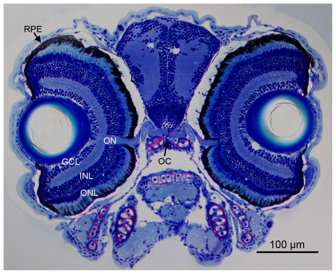
Retina morphology of a 6-day-old larva. Histology shows the vertebrate typical layering of the larval zebrafish retina with the optic nerve and chiasm. ONL, outer nuclear layer; INL, inner nuclear layer; GLC, ganglion cell layer; ON, optic nerve; RPE, retinal pigment epithelium; OC, optic chiasm.
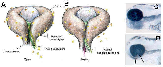
(A,B) Schematics of the forming eye showing the ventrally positioned choroid fissure before and during closure (drawing by Clarissa Scholes). (C,D) Wildtype (C) and colobomatous (D) zebrafish eyes
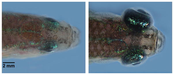
Dorsal views of wild-type (Left) and bugeye mutant (Right) adult zebrafish.
References
-
- Adamis AP, Aiello LP, D’Amato RA. Angiogenesis and ophthalmic disease. Angiogenesis. 1999;3:9–14. - PubMed
-
- Agathocleous M, Harris WA. From progenitors to differentiated cells in the vertebrate retina. Annu Rev Cell Dev Biol. 2009;25:45–69. - PubMed
-
- Akhtar S, Schonthaler HB, Bron AJ, Dahm R. Formation of stromal collagen fibrils and proteoglycans in the developing zebrafish cornea. Acta Ophthalmol. 2008;86:655–665. - PubMed
-
- Allaman I, Belanger M, Magistretti PJ. Astrocyte-neuron metabolic relationships: for better and for worse. Trends in Neuroscience. 2011;34:76–87. - PubMed
