Review
. 2011 May 19:9:53.
doi: 10.1186/1477-7819-9-53.

Extracranial propagation of glioblastoma with extension to pterygomaxillar fossa

Affiliations
Review

Extracranial propagation of glioblastoma with extension to pterygomaxillar fossa

Damir Tomac et al. World J Surg Oncol. .

Abstract

Background: Glioblastoma multiforme is a highly malignant primary brain tumor that shows marked local aggressiveness, but extracranial spread is not a common occurrence. We present an unusual case of recurrent glioblastoma in 54-year old male that spread through the scull base to the ethmoid and sphenoid sinuses, to the orbita, pterygomaxillar fossa, and to the neck.

Methods: A 54-year old male underwent left temporal resection because of brain tumor of his left temporal lobe. Operation was followed by external beam radiation combined with temozolomide. The tumor recurred eight months after first surgery. The patient developed swelling of left temporal region, difficult swallowing and headache. MRI of head showed recurrent tumor, which invaded orbita, ethmoid and sphenoid sinuses, nasal cavity, pterygomaxillar fossa.

Results: The patient died ten months after initial diagnosis of glioblastoma multiforme, and two months after his second operation.

Conclusions: The aggressive surgical operation helped to downsize the tumor mass as much as possible, but did not prolonged significantly the life or improved the life quality of the patient. The current literature is reviewed, and the diagnostic approaches as well as therapeutic options are discussed.


Figures
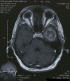
Figure 1
Figure 1
Axial T1 weighted contrast enhanced MRI image demonstrating ring-enhanced lesion of the left temporal lobe
Figure 2
Figure 2
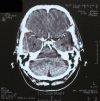
Axial contrast enhanced MSCT of the head showing no signs of tumor.
Figure 3
Figure 3
Axial T1-weighted contrast enhanced MRI image demonstrates extra cranial portion of the tumor extended to the sphenoid and ethmoid sinuses, nasal cavity, and orbit.
Figure 4
Figure 4
Sagittal T1-weighted contrast enhanced MRI image demonstrates intracranial and extra cranial portion of the tumor extended to the sphenoid and ethmoid sinuses, nasal cavity, orbit, pterygomaxillar fossa, and neck.
Figure 5
Figure 5
Tissue sections showing a glioblastoma multiforme with invasion in muscle tissue (hematoxylin-eosin stain, ×400).
Figure 6
Figure 6
Tissue sections showing a glioblastoma multiforme with bone invasion (hematoxylin-eosin stain, ×400)
Figure 7
Figure 7
Tissue sections showing a glioblastoma multiforme with angioinvasion (hematoxylin-eosin stain, ×400).

References

    1. Taha M, Ahmad A, Wharton S, Jellinek D. Extra-cranial metastasis of glioblastoma multiforme presenting as acute parotitis. Br J Neurosurg. 2005;19:348–351. doi: 10.1080/02688690500305506. - DOI - PubMed
    1. Pasquier B, Pasquier D, N'Golet A, Panh MH, Couderc P. Extraneural metastases of astrocytomas and glioblastomas: clinicopathological study of two cases and review of literature. Cancer. 1980;45:112–125. doi: 10.1002/1097-0142(19800101)45:1<112::AID-CNCR2820450121>3.0.CO;2-9. - DOI - PubMed
    1. Ates LE, Bayindir C, Bilgic B, Karasu A. Glioblastoma with lymph node metastases. Neuropathology. 2003;23:146–149. doi: 10.1046/j.1440-1789.2003.00490.x. - DOI - PubMed
    1. Wallace CJ, Forsyth PA, Edwards DR. Lymph node metastases from glioblastoma multiforme. AJNR Am J Neuroradiol. 1996;17:1929–1931. - PMC - PubMed
    1. Rubinstein LJ. Development of extracranial metastases from a malignant astrocytoma in the absence of previous craniotomy. Case report. J Neurosurg. 1967;26:542–547. doi: 10.3171/jns.1967.26.5.0542. - DOI - PubMed