. 2011 May 19:10:37.
doi: 10.1186/1475-2859-10-37.

Overexpression of human virus surface glycoprotein precursors induces cytosolic unfolded protein response in Saccharomyces cerevisiae

Affiliations

Overexpression of human virus surface glycoprotein precursors induces cytosolic unfolded protein response in Saccharomyces cerevisiae

Evaldas Ciplys et al. Microb Cell Fact. .

Abstract

Background: The expression of human virus surface proteins, as well as other mammalian glycoproteins, is much more efficient in cells of higher eukaryotes rather than yeasts. The limitations to high-level expression of active viral surface glycoproteins in yeast are not well understood. To identify possible bottlenecks we performed a detailed study on overexpression of recombinant mumps hemagglutinin-neuraminidase (MuHN) and measles hemagglutinin (MeH) in yeast Saccharomyces cerevisiae, combining the analysis of recombinant proteins with a proteomic approach.

Results: Overexpressed recombinant MuHN and MeH proteins were present in large aggregates, were inactive and totally insoluble under native conditions. Moreover, the majority of recombinant protein was found in immature form of non-glycosylated precursors. Fractionation of yeast lysates revealed that the core of viral surface protein aggregates consists of MuHN or MeH disulfide-linked multimers involving eukaryotic translation elongation factor 1A (eEF1A) and is closely associated with small heat shock proteins (sHsps) that can be removed only under denaturing conditions. Complexes of large Hsps seem to be bound to aggregate core peripherally as they can be easily removed at high salt concentrations. Proteomic analysis revealed that the accumulation of unglycosylated viral protein precursors results in specific cytosolic unfolded protein response (UPR-Cyto) in yeast cells, characterized by different action and regulation of small Hsps versus large chaperones of Hsp70, Hsp90 and Hsp110 families. In contrast to most environmental stresses, in the response to synthesis of recombinant MuHN and MeH, only the large Hsps were upregulated whereas sHsps were not. Interestingly, the amount of eEF1A was also increased during this stress response.

Conclusions: Inefficient translocation of MuHN and MeH precursors through ER membrane is a bottleneck for high-level expression in yeast. Overexpression of these recombinant proteins induces the UPR's cytosolic counterpart, the UPR-Cyto, which represent a subset of proteins involved in the heat-shock response. The involvement of eEF1A may explain the mechanism by which only large chaperones, but not small Hsps are upregulated during this stress response. Our study highlights important differences between viral surface protein expression in yeast and mammalian cells at the first stage of secretory pathway.


Figures

Figure 1
Figure 1
Synthesis of recombinant MuHN and MeH causes overexpression of some cellular proteins in yeast. (A-C) SDS-PAGE of whole cell lysates on a 10% polyacrylamide gel. All lysates were prepared from galactose-induced yeast cells of S. cerevisiae pep4 strain, transformed with empty vector (lane 1) or plasmids expressing MuHN (native sequence - lane 2 and with His6-tag - lane 3) or His6-tagged MeH (lane 4). Lane M - prestained protein ladder. (A) Coomassie blue-stained gel. Solid arrows indicate bands of recombinant MuHN (lanes 2 and 3) and MeH (lane 4) proteins. Dashed arrow points to ~70 kDa main band of yeast cellular proteins overexpressed in response to synthesis of MuHN and MeH (lanes 2-4). (B) Western blot using anti-His antibody. Expression of His-tagged recombinant MeH protein was confirmed by immunoblot analysis that revealed two main forms: major band of ~65 kDa and upper double band of ~75 kDa (lane 4, there are also a faint band just above 65 kDa and some degradation products visible). His-tagged recombinant MuHN protein did not react with anti-His antibody (lane 3). (C) Western blot using monoclonal antibody 782 to the native MuHN. Both native and His-tagged MuHN sequence variants were detected as ~60 kDa bands (lanes 2 and 3, respectively) corresponding to those shown in coomassie stained gel. Due to stronger reaction of Mab 782 with the native sequence MuHN variant the latter was chosen for further MuHN expression study. There was also some non-specific reaction and cross-reactivity with MeH observed.
Figure 2
Figure 2
SDS-PAGE of recombinant MuHN and MeH protein fractions insoluble in 8 M urea solution. (A-D) The same samples were run on each gel: control sample from S. cerevisiae cells, transformed with empty vector was loaded on lane 1, whereas samples from S. cerevisiae expressing recombinant viral proteins were loaded on lanes 2 (MuHN) and 3 (MeH), respectively. (A) Coomassie blue-stained gel. Long solid arrows indicate major, whereas dotted arrows - minor forms of partially purified recombinant MuHN (lane 2) and MeH (lane 3). Short arrow points to ~50 kDa band analysed by MS directly from 1-D SDS-PAGE gel (the band number 15 is given according to the list of identified yeast proteins in Table 1). (B) Western blot using monoclonal antibody 782 to the native MuHN. In addition to the main band of ~60 kDa (solid arrow) the minor band (up to 65 kDa, dotted arrow) of MuHN was also distinguished (lane 2). Mab 782 cross-reacted with both major and minor bands of recombinant MeH (lane 3). (C) Western blot using anti-His antibody. Both ~65 kDa and ~75 kDa forms of MeH protein (indicated by solid and dotted arrows, respectively) were insoluble under mild denaturing conditions in 8 M urea solution (lane 3). (D) Western blot using Concanavalin A. The major forms of recombinant MuHN (~60 kDa) and MeH (~65 kDa) appeared to be non-glycosylated as they did not react with Concanavalin A (white band areas below dotted arrows in lanes 2 and 3, respectively). Only heterogeneous band of minor MeH form (~75 kDa) contained N-glycosylated protein reacting with Concanavalin A (lane 3, dotted arrow), similar result was observed in the case of MuHN minor form (lane 2).
Figure 3
Figure 3
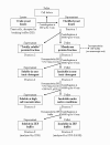
A flow diagram for fractionation of yeast lysates.
Figure 4
Figure 4
PNGase F treatment of S. cerevisiae whole cell lysates followed by Western blotting using anti-His antibody. Lysates were prepared from galactose-induced yeast cells, transformed with either an empty vector (lane C) or plasmids expressing the full sequence MeH variant (pFGG3-MeH) and chimeric α-MeH protein with α-factor signal sequence instead of native N-terminal TM domain (pFGG3-alpha-MeH). Lysates with (+) or without (-) PNGase F treatment are indicated below blot lanes. Solid arrows indicate unglycosylated MeH polypeptides, dotted arrows - glycosylated MeH forms.
Figure 5
Figure 5
Expression of His-tagged MeH in P. pastoris transformants induced with methanol. (A) SDS-PAGE of whole cell lysates from P. pastoris transformants carrying empty vector or multicopy MeH expression cassetes. (B) Western blotting of the same samples using anti-tetraHis antibody. M- prestained protein marker, C- Control (pPIC3.5K MutS), 1,2,3- multicopy pPIC3.5K-MeH transformants for intracellular expression of MeH, resistant to various concentrations of antibiotic G418. Solid arrows indicate unglycosylated MeH polypeptide precursor, dotted arrows point to glycosylated MeH form and dashed arrows show cellular proteins, upregulated in response to MeH expression.
Figure 6
Figure 6
Analysis of MeH in insoluble protein fractions from P. pastoris by Western blotting using anti-His antibody. The fractions insoluble in 1% TritonX-100 and 1M NaCl were obtained from P. pastoris transformants using the same method as described in Methods for S. cerevisiae samples (correspond to fraction 7 in Figure 3). VK indicates the sample obtained from P. pastoris transformant carrying one copy of pPIC3.5K-MeH expression cassete. Other markings are the same as in Figure 5 (dotted arrow indicates glycosylated MeH form, whereas solid arrow - unglycosylated MeH precursor).
Figure 7
Figure 7
Analysis of the expression of α-MeH chimeric protein containing S. cerevisiae α-factor signal sequence. Whole cell lysates of methanol induced P. pastoris cells (Mut+ phenotype) expressing α-MeH chimeric protein containing S. cerevisiae α-factor signal sequence instead of native TM anchor domain were resolved by SDS-PAGE, blotted onto nitrocellulose membrane and analysed by Western blotting using anti-His antibody. M- protein marker, C- control (pPIC9K Mut+), VK- one-copy pPIC9K-MeH transformant, 1,2,3- multicopy pPIC9K-MeH transformants. Solid arrow indicates unglycosylated MeH polypeptides, dotted arrow - glycosylated MeH forms.
Figure 8
Figure 8
Analysis of MeH expression in whole cell lysates from P. pastoris and S. cerevisiae. Whole cell lysates from P. pastoris (C, In, SP and SG) and S. cerevisiae (Sc) were analysed by Western blotting using anti-His antibody. M- marker, C- control, In- multicopy pPIC3.5K-MeH transformant for intracellular expression of full sequence MeH protein, SP- multicopy pPIC9K-MeH transformant for secreted expression of chimeric α-MeH protein with α-factor signal sequence (overexpressing both polypeptide precursor and glycosylated MeH forms), SG- another multicopy pPIC9K-MeH transformant for secreted expression of chimeric α-MeH, overexpressing glycosylated MeH form, Sc- pFGG3-MeH transformant of S. cerevisiae AH22 strain overexpressing full sequence MeH protein after induction with galactose. Solid arrows indicate unglycosylated MeH polypeptides, whereas dotted arrows - glycosylated MeH forms.
Figure 9
Figure 9
Recombinant MuHN and MeH induce specific stress response in yeast cells. (A-I) 2D gel electrophoresis of yeast proteins. Samples were taken from yeast cells of S. cerevisiae AH22 strain, expressing MuHN (central panel; B-H) or MeH (right panel; C-I) and from control AH22 cells, transformed with empty vector pFGG3 (left panel; A-G). At the top (A-C) whole cell lysates, in the middle (D-F) fractions of proteins, soluble at high salt concentration, and in the bottom panel (G-I) proteins, insoluble under native conditions are shown. Solid arrows in B and C indicate proteins, identified by MS directly from whole cell lysates, whereas dotted arrows point to the proteins, identified from soluble and insoluble fractions. Numbers of identified protein spots correspond to those, given in Table 1. Protein molecular mass markers (120, 66, 45, 35, and 25 kDa) were run simultaneously in the left lane (M) of each 2D gel.

References

    1. Mattanovich D, Gasser B, Hohenblum H, Sauer M. Stress in recombinant protein producing yeasts. J Biotechnol. 2004;113:121–135. doi: 10.1016/j.jbiotec.2004.04.035. - DOI - PubMed
    1. Gasser B, Saloheimo M, Rinas U, Dragosits M, Rodríguez-Carmona E, Baumann K, Giuliani M, Parrilli E, Branduardi P, Lang C, Porro D, Ferrer P, Tutino ML, Mattanovich D, Villaverde A. Protein folding and conformational stress in microbial cells producing recombinant proteins: a host comparative overview. Microb Cell Fact. 2008;7:11. doi: 10.1186/1475-2859-7-11. - DOI - PMC - PubMed
    1. Freigassner M, Pichler H, Glieder A. Tuning microbial hosts for membrane protein production. Microb Cell Fact. 2009;8:69. doi: 10.1186/1475-2859-8-69. - DOI - PMC - PubMed
    1. Metzger MB, Michaelis S. Analysis of quality control substrates in distinct cellular compartments reveals a unique role for Rpn4p in tolerating misfolded membrane proteins. Mol Biol Cell. 2009;20:1006–1019. - PMC - PubMed
    1. Geiler-Samerotte KA, Dion MF, Budnik BA, Wang SM, Hartl DL, Drummond DA. Misfolded proteins impose a dosage-dependent fitness cost and trigger a cytosolic unfolded protein response in yeast. Proc Natl Acad Sci USA. 2011;108:680–685. doi: 10.1073/pnas.1017570108. - DOI - PMC - PubMed

Publication types

MeSH terms

LinkOut - more resources