. 2011 May 20;30(1):62.
doi: 10.1186/1756-9966-30-62.

Increased IL-10 mRNA expression in tumor-associated macrophage correlated with late stage of lung cancer

Affiliations

Increased IL-10 mRNA expression in tumor-associated macrophage correlated with late stage of lung cancer

Rui Wang et al. J Exp Clin Cancer Res. .

Abstract

Background: Monocyte recruited into the tumor and maturation to tumor-associated macrophage (TAM). Interleukin-10(IL-10) is a potent immunosuppressive cytokine, which can be secreted from both primary tumor and stromal cells. However, there are controversies regarding its role in the progression of cancer. So it is important to isolate TAM from tumor cells to study the role of IL-10 in the progress of cancer. The aim of our study was to determine whether IL-10 expressed by TAM correlated with clinicopathological factors in NSCLC.

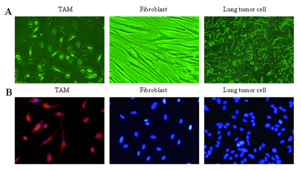
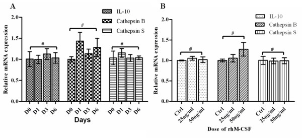
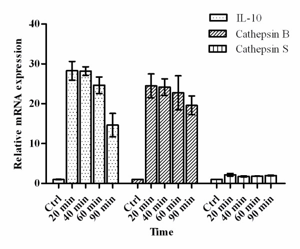
Methods: TAM in NSCLC was isolated by short-term culture in serum free medium with the modification to literature reports. The mRNA expression levels of IL-10, cathepsin B, cathepsin S, which were closely related with TAM according to the literatures, were evaluated by Quantitative real-time RT-PCR in 63 NSCLC. The relationships between their expression levels and clinicopathological features were investigated.

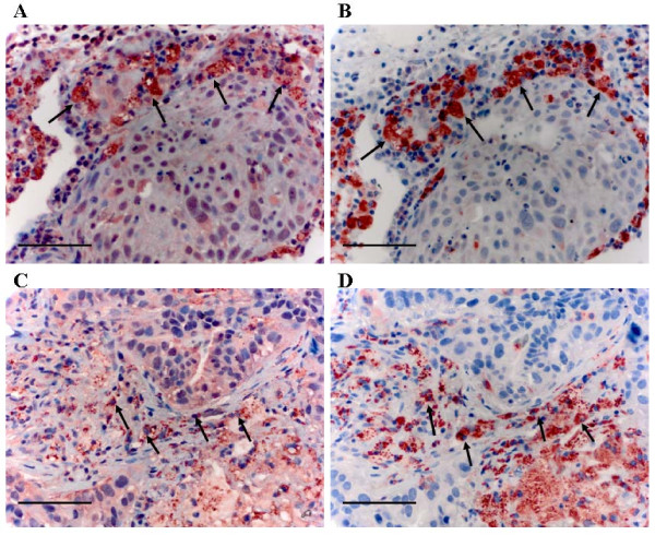
Results: We successfully achieved up to 95% purity of TAM, derived from 63 primary lung cancer tissues. TAM expressed high levels of IL-10, cathepsin B in NSCLC. High levels of IL-10 in TAM significantly correlated with stage, tumor size, lymph node metastasis, lymphovascular invasion or histologic poor differentiation.

Conclusions: Our results revealed that TAM with high levels of IL-10 expression may play an important role in the progression of non-small cell lung cancer. The data also suggested that TAMs may involve in tumor immunosuppression through overexpressed IL-10. Additionally, the phenotype of isolated TAM can be potentially used to predict clinicopathological features as well.


Figures

Figure 1
Figure 1
Characterization of tumor-associated macrophage. A. Representative cell morphology of tumor-associated macrophages, TAM, fibroblast and lung tumor cell. B. Immunofluorescent was used to distinguish macrophage, fibroblast and lung tumor cell with antibodies targeting CD68 (red), nuclei stained with DAPI (blue). Original magnification, × 400.
Figure 2
Figure 2
The mRNA expression levels of IL-10, cathepsin B and cathepsin S in normal macrophages. Results are given as fold increase in mRNA expression with respect to expression in D0 monocytes. Data were normalized to expression of the β-actin gene. A: Monocytes(D0) was used as a calibrator. B, monocytes culture without rhM-CSF was used as a calibrator (Ctrl). Error bar is SD, Independent experiments were repeated three times, all #p > 0.05(by student t-test).
Figure 3
Figure 3
The mRNA expression levels of IL-10, cathepsin B and cathepsin S in TAM changes in primary culture. Results are given as fold increase in mRNA expression with respect to expression in ctrl (normal macrophages). Data were normalized to expression of the β-actin gene. Normal macrophages were used as a calibrator. Error bar is SD; Independent experiments were repeated three times.
Figure 4
Figure 4
mRNA from TAMs and matched normal macrophage(Mφ) was analyzed by Quantitative real-time RT-PCR for expression of the indicated genes in 63 NSCLC samples. Results are given as fold increase in mRNA expression with respect to expression in matched Mφ. Data were normalized to expression of the β-actin gene. Mφ was used as a calibrator. Bars represent median. *p by the Mann-Whitney U test.
Figure 5
Figure 5
Immunohistochemical expression of IL-10, cathepsin B and CD68 in macrophage. A-B, High IL-10 expression in macrophage, A, IL-10 staining in macrophage (strong positivity); B, CD68 staining. C-D, Cathepsin B expression in macrophage; C, cathepsin B staining in macrophage (most cells were moderate positivity, only a few cells were strong staining); D, CD68 staining. Scale bar indicates 50 μm. Original magnification, × 400.

References

    1. Pollard JW. Tumour-educated macrophages promote tumour progression and metastasis. Nat Rev Cancer. 2004;4(1):71–78. doi: 10.1038/nrc1256. - DOI - PubMed
    1. Balkwill F, Mantovani A. Inflammation and cancer: back to Virchow? Lancet. 2001;357(9255):539–545. doi: 10.1016/S0140-6736(00)04046-0. - DOI - PubMed
    1. Joyce JA, Pollard JW. Microenvironmental regulation of metastasis. Nat Rev Cancer. 2009;9(4):239–252. doi: 10.1038/nrc2618. - DOI - PMC - PubMed
    1. Ohno S, Ohno Y, Suzuki N, Kamei T, Koike K, Inagawa H, Kohchi C, Soma G, Inoue M. Correlation of histological localization of tumor-associated macrophages with clinicopathological features in endometrial cancer. Anticancer Res. 2004;24(5C):3335–3342. - PubMed
    1. Takanami I, Takeuchi K, Kodaira S. Tumor-associated macrophage infiltration in pulmonary adenocarcinoma: association with angiogenesis and poor prognosis. Oncology. 1999;57(2):138–142. doi: 10.1159/000012021. - DOI - PubMed

Publication types