. 2011 Aug;230(2):258-72.
doi: 10.1016/j.expneurol.2011.05.004. Epub 2011 May 8.

Some lumbar sympathetic neurons develop a glutamatergic phenotype after peripheral axotomy with a note on VGLUT₂-positive perineuronal baskets

Affiliations

Some lumbar sympathetic neurons develop a glutamatergic phenotype after peripheral axotomy with a note on VGLUT₂-positive perineuronal baskets

Pablo R Brumovsky et al. Exp Neurol. 2011 Aug.

Abstract

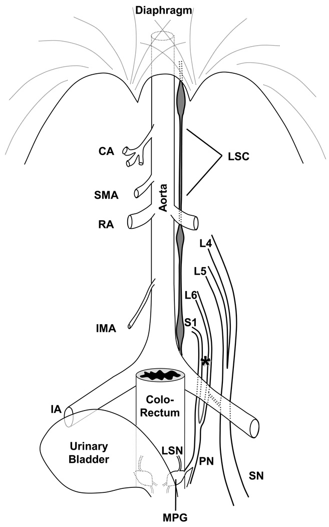
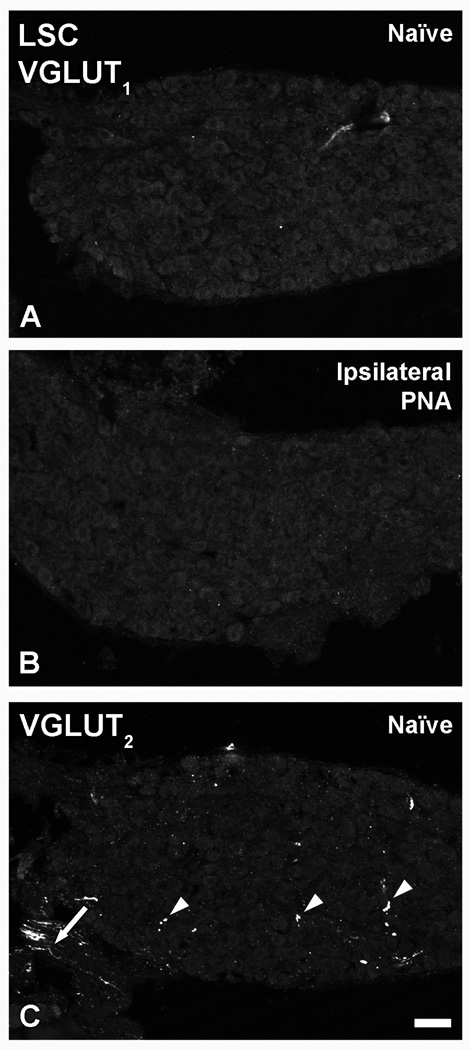
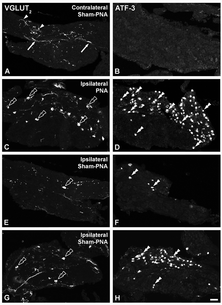
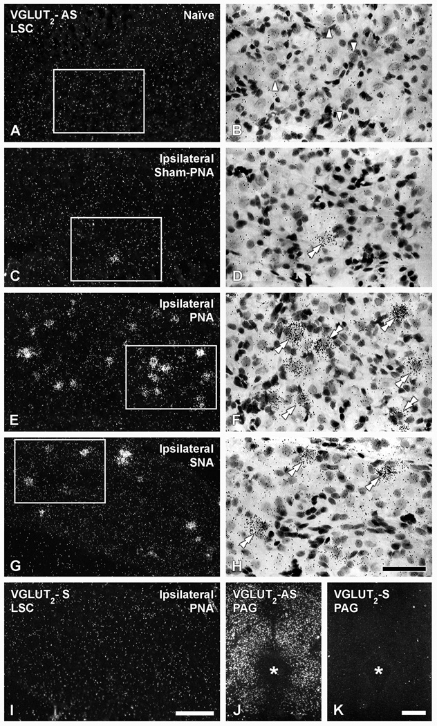
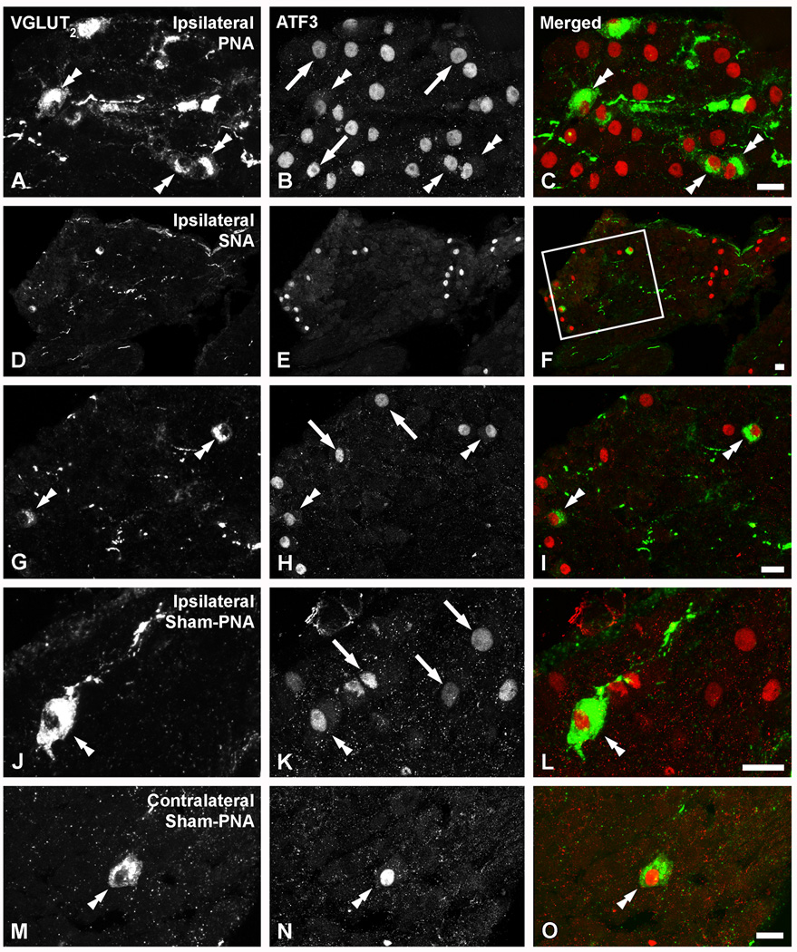
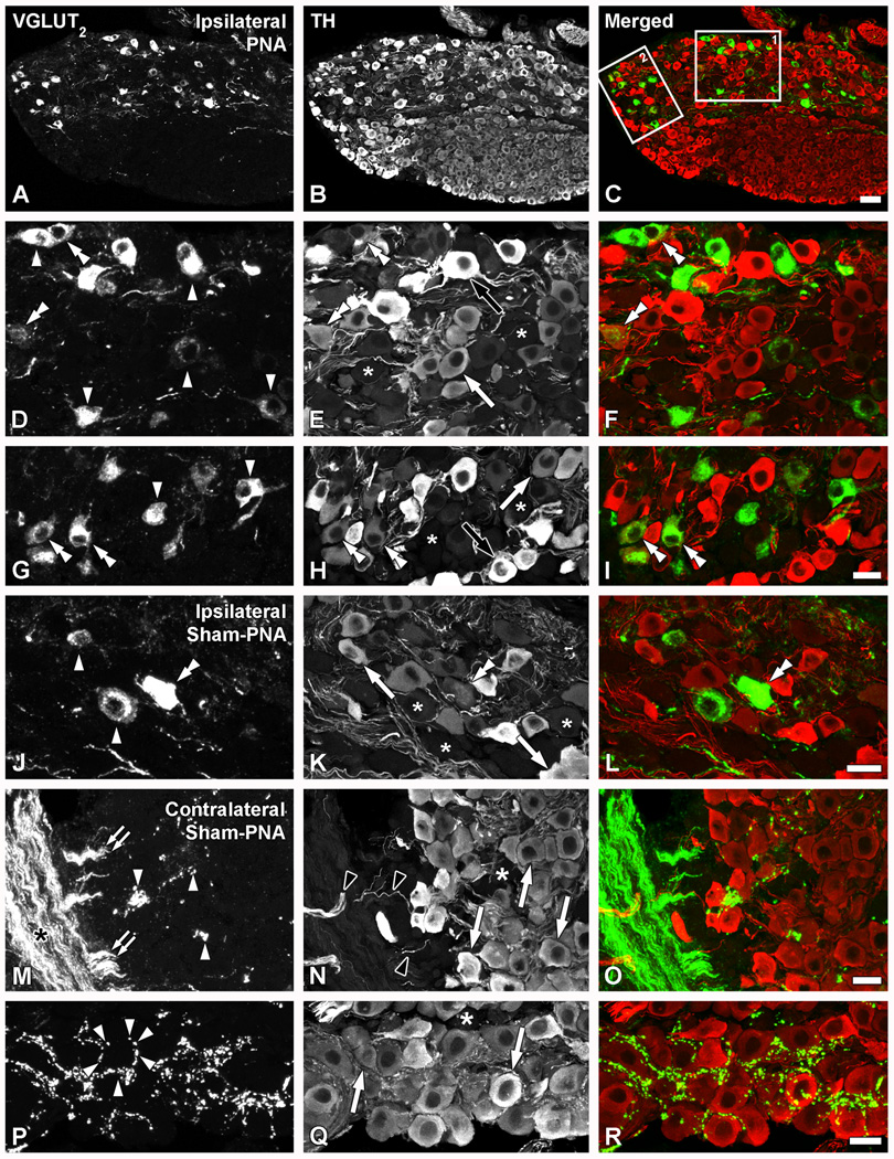
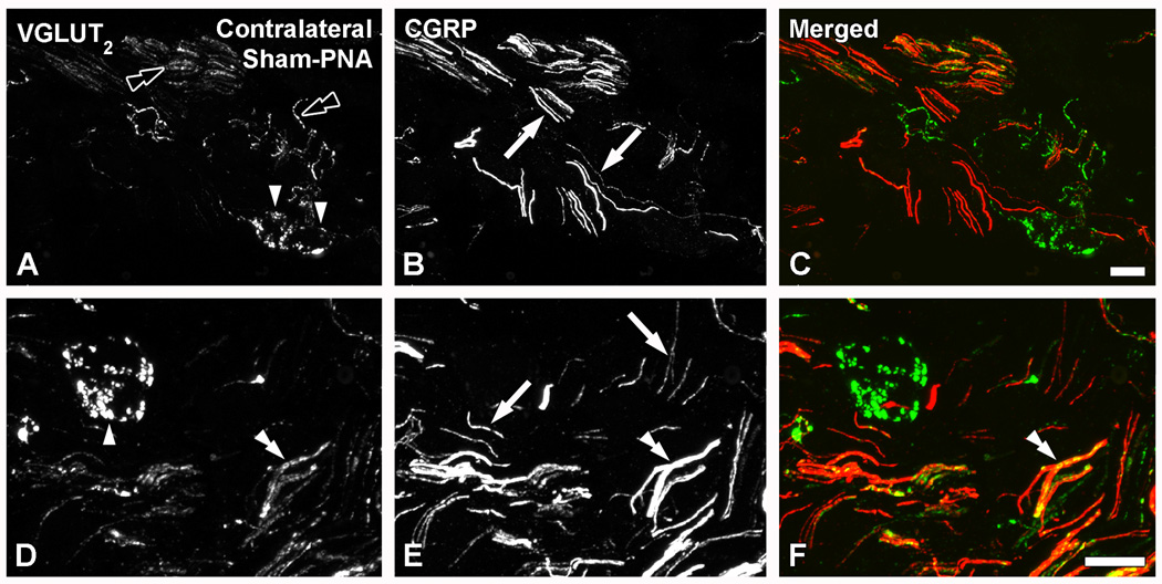
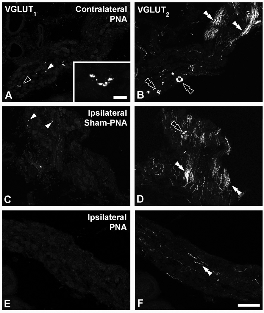
Glutamate is the main excitatory neurotransmitter in the nervous system, including in primary afferent neurons. However, to date a glutamatergic phenotype of autonomic neurons has not been described. Therefore, we explored the expression of vesicular glutamate transporter (VGLUT) types 1, 2 and 3 in lumbar sympathetic chain (LSC) and major pelvic ganglion (MPG) of naïve BALB/C mice, as well as after pelvic nerve axotomy (PNA), using immunohistochemistry and in situ hybridization. Colocalization with activating transcription factor-3 (ATF-3), tyrosine hydroxylase (TH), vesicular acetylcholine transporter (VAChT) and calcitonin gene-related peptide was also examined. Sham-PNA, sciatic nerve axotomy (SNA) or naïve mice were included. In naïve mice, VGLUT(2)-like immunoreactivity (LI) was only detected in fibers and varicosities in LSC and MPG; no ATF-3-immunoreactive (IR) neurons were visible. In contrast, PNA induced upregulation of VGLUT(2) protein and transcript, as well as of ATF-3-LI in subpopulations of LSC neurons. Interestingly, VGLUT(2)-IR LSC neurons coexpressed ATF-3, and often lacked the noradrenergic marker TH. SNA only increased VGLUT(2) protein and transcript in scattered LSC neurons. Neither PNA nor SNA upregulated VGLUT(2) in MPG neurons. We also found perineuronal baskets immunoreactive either for VGLUT(2) or the acetylcholinergic marker VAChT in non-PNA MPGs, usually around TH-IR neurons. VGLUT(1)-LI was restricted to some varicosities in MPGs, was absent in LSCs, and remained largely unaffected by PNA or SNA. This was confirmed by the lack of expression of VGLUT(1) or VGLUT(3) mRNAs in LSCs, even after PNA or SNA. Taken together, axotomy of visceral and non-visceral nerves results in a glutamatergic phenotype of some LSC neurons. In addition, we show previously non-described MPG perineuronal glutamatergic baskets.


Figures

Figure 1
Figure 1
Schematic diagram showing the general anatomical distribution of the lumbar sympathetic chain (LSC), the ventral branches of the spinal nerves L4-L6 and S1, the major pelvic ganglion (MPG), and the pelvic (PN), lumbar splanchnic (LSN) and sciatic (SN) nerves in the male mouse. Their relation with the aorta and stemming branches (celiac artery (CA), superior mesenteric artery (SMA), renal artery (RA), inferior mesenteric artery (IMA), iliac artery (IA)) is emphasized. Not illustrated for clarity is the spine, spinal cord or dorsal root ganglia, from which the axons of the spinal nerves arise. The nerves were exposed by blunt dissection of the overlying lumbar muscles (not shown); the location where transection of the pelvic nerve tributaries was executed is shown with an asterisk. The MPG is located between the colorectum and the urinary bladder, with branches from the pelvic nerve traveling through it (for clarity, other branches entering or exiting the MPG are not illustrated).
Figure 2
Figure 2
Confocal immunofluorescence photomicrographs of sections of the LSC from naïve mice (A, C) or PNA mice (B), incubated with VGLUT1 (A, B) or VGLUT2 (C) antisera. Arrowheads and arrow in (C) show VGLUT2-IR varicosities and nerve fibers, respectively in naïve LSCs. Scale bar: 50 µm.
Figure 3
Figure 3
Confocal immunofluorescence photomicrographs of sections of the contralateral (A, B) or ipsilateral (C–H) LSC of mice 7 days after sham-PNA (A,B; E–H) or PNA (C, D), incubated with VGLUT2 (A, C, E, G) or ATF-3 (B, D, F, H) antisera (each corresponding pair of VGLUT2 or ATF-3 micrographs shows sections separated by 36 µm). Arrows and arrowheads in (A, B) show VGLUT2-IR fibers and varicosities, respectively. Black double arrowheads and double arrowheads in (C, D, E–H) show VGLUT2- and ATF-3-IR LSC NPs. Scale bar: 50 µm (H=A–G).
Figure 4
Figure 4
Dark-(A, C, E, G, I, J, K) and bright-field (B, D, F, H) photomicrographs of LSC (A–H; I) or the PAG (J, K), in naïve (A, B, J, K), ipsilateral sham-PNA (C, D), ipsilateral PNA (E, F, I) or ipsilateral SNA (G, H), after hybridization with antisense (A–H; J) and sense (I, K) riboprobes for VGLUT2 mRNA (B, D, F and H show high power magnification images of boxes in A, C, E and G, respectively). (Arrowheads in B show NPs lacking VGLUT2 mRNA. Double arrowheads in D, F, H show VGLUT2 mRNA-expressing NPs. Asterisk shows location of the aqueduct of Sylvius. Scale bars: 50 µm (H=B, D, F); 100 µm (I=A, C, E, G); 500 µm (J, K).
Figure 5
Figure 5
Dark-field photomicrographs of LSC (A–H), the hippocampus (HC) (I, J) or the dorsal raphe nucleus (DR) (K, L), in naïve (A, B, I–L), ipsilateral sham-PNA (C, D), ipsilateral PNA (E, F) or ipsilateral SNA (G, H), after hybridization with antisense (A–H; I, K) and sense (J, L) riboprobes for VGLUT1 (A, C, E, G, I, J) or VGLUT3 (B, D, F, H, K, L) mRNAs. Double arrow in (I) and double arrows in (K) show mRNA labeling in the CA3 region of the HC or the DR, respectively. Scale bars: 50 µm (H=A–G); 200 µm (K, L); 500 µm (I, J).
Figure 6
Figure 6
Confocal immunofluorescence photomicrographs of sections of the ipsilateral LSC of mice 7 days after PNA (A–C), 10 days after SNA (D–I), ipsilateral (J–L) or contralateral to a sham-PNA (M–O), incubated with VGLUT2 (A, D, G, J, M) and ATF-3 (B, E, H, K, N) antisera (C, F, I, L, O show merged images). (D–I; G–H show a magnified view of the box in F). Double arrowheads in (A, G, J, M, B, H, K, N, C, I, L, O) and arrows in (B, H, K) show VGLUT2/ATF-3 or ATF-3-only neurons, respectively. Scale bars: 20 µm (C=A, B; F=D, E; I=G, H; L=J, K; O=M, N).
Figure 7
Figure 7
Confocal immunofluorescence photomicrographs of sections of the ipsilateral (A–L) or contralateral (M–R) LSCs of mice 7 days after PNA (A–I) or sham-PNA (J–R), incubated with VGLUT2 (A, D, G, J, M, P) and TH (B, E, H, K, N, Q) antisera (C, F, I, L, O, R show merged images) (asterisks in E, H, K, N, Q indicate TH-negative LSC NPs). (A–I; boxes 1 and 2 in C are shown at higher magnification in D–F and G–I, respectively). Arrowheads in (D, G, J) and double arrowheads in (D, G, J, B, H, K, F, I, L) show TH-only or VGLUT2/TH neurons, respectively. White arrows and black arrows show light or strong TH-IR-only NPs, respectively (M–R). Black asterisk in M shows thick, VGLUT2-IR fiber bundles, from which smaller bundles appear to detach (double arrowheads in M). Black arrowheads show TH-IR fibers. Perineuronal VGLUT2-IR baskets (arrowheads in P) are occasionally seen around TH-IR NPs (arrows in Q). Scale bars: 50 µm (C=A, B); 20 µm (I= D–H; L=J, K; O=M, N; R=P, Q).
Figure 8
Figure 8
Confocal immunofluorescence photomicrographs of sections of the contralateral MPGs of sham-PNA mice, co-incubated with VGLUT2 (A, D) and CGRP (B, E) antisera (C, F, show merged images). Black double arrowheads in (A) and arrowheads in (A, D) show VGLUT2-IR nerves or perineuronal baskets, respectively. Arrows in (B, E) and double arrowhead in (D–F) show CGRP fibers or VGLUT2/CGRP-double stained nerve bundles. Scale bars: 20 µm (C=A, B; F=D, E).
Figure 9
Figure 9
Confocal immunofluorescence photomicrographs of sections from contralateral (A–D; M–P) or ipsilateral (E–L) MPGs after PNA (A–D; M–P) or sham-PNA (E–L), incubated with VGLUT2 (A, E, I, M), VAChT (B, F, J, N) and TH (C, G, K, O) antisera (D, H, L, P, show merged images). Arrowheads in (A, E, I) and white arrows in (B, F, J) show two separate populations of TH-IR neurons showing VGLUT2- or VAChT-IR perineuronal baskets, respectively. Double arrowhead in (A) and black double arrowhead in (C) show thick VGLUT2- or TH-IR nerve bundles, respectively. Black arrows in (M–P) show some TH-IR NPs innervated by both VGLUT2- and VAChT-IR baskets. Scale bars: 50 µm (D=A–C); 10 µm (H=E–G; L=I–K; P=M–O).
Figure 10
Figure 10
Confocal immunofluorescence photomicrographs of sections from contralateral (A, B) or ipsilateral (C–F) MPGs after PNA (A, B, E, F) or sham-PNA (c, D), incubated with VGLUT1 (A, C, E) or VGLUT2 (B, D, F) antisera. White arrowheads in (A, C) show VGLUT1-IR varicosities (shown at higher magnification in inset (black arrowhead)). White or black double arrowheads show nerve fibers (B, D, F) or perineuronal baskets (B, D), respectively. Scale bars: 100 µm (A–F); 10 µm (inset in A).

References

    1. Adams JC. Biotin amplification of biotin and horseradish peroxidase signals in histochemical stains. J. Histochem. Cytochem. 1992;40:1457–1463. - PubMed
    1. Aïoun J, Rampin O. Anatomical evidence for glutamatergic transmission in primary sensory neurons and onto postganglionic neurons controlling penile erection in rats: an ultrastructural study with neuronal tracing and immunocytochemistry. Cell. Tissue Res. 2006;323:359–375. - PubMed
    1. Apostolova G, Dechant G. Development of neurotransmitter phenotypes in sympathetic neurons. Auton. Neurosci. 2009;151:30–38. - PubMed
    1. Balazs R. Trophic effect of glutamate. Curr. Top. Med. Chem. 2006;6:961–968. - PubMed
    1. Bennett GJ, Xie YK. A peripheral mononeuropathy in rat that produces disorders of pain sensation like those seen in man. Pain. 1988;33:87–107. - PubMed

Publication types

Substances