. 2011 Jul;141(1):176-85.
doi: 10.1053/j.gastro.2011.04.010. Epub 2011 Apr 16.

MyD88 and retinoic acid signaling pathways interact to modulate gastrointestinal activities of dendritic cells

Affiliations

MyD88 and retinoic acid signaling pathways interact to modulate gastrointestinal activities of dendritic cells

Eduardo J Villablanca et al. Gastroenterology. 2011 Jul.

Abstract

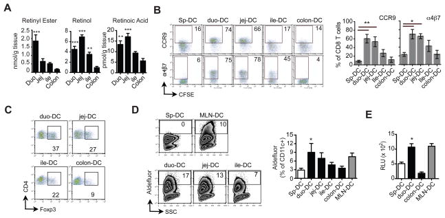
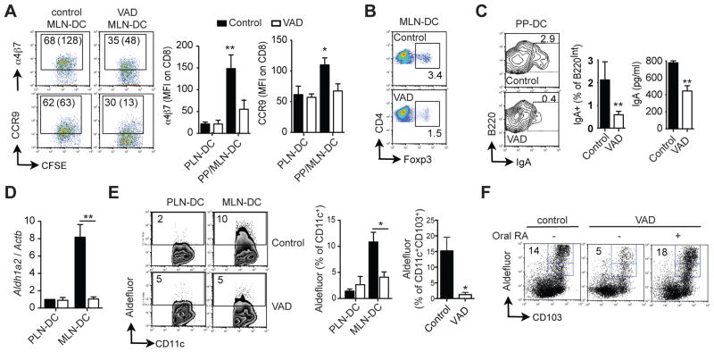
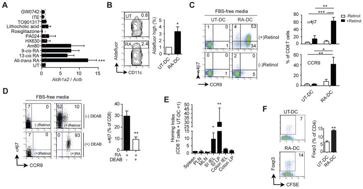
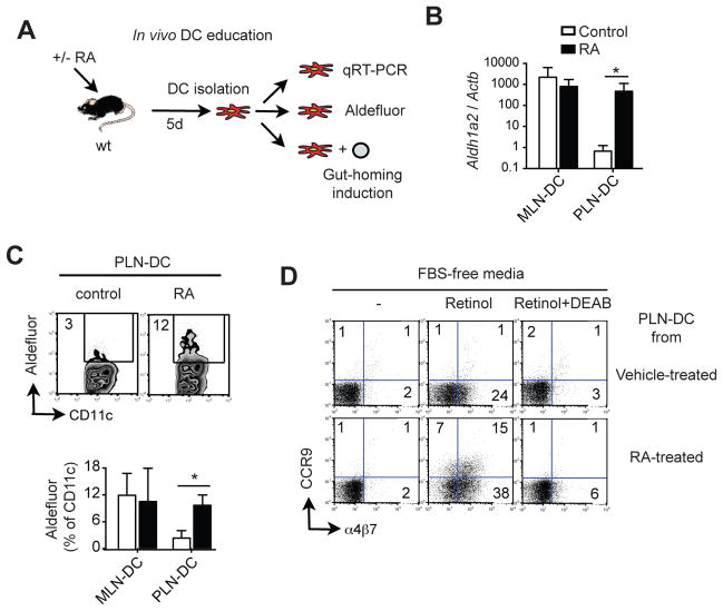
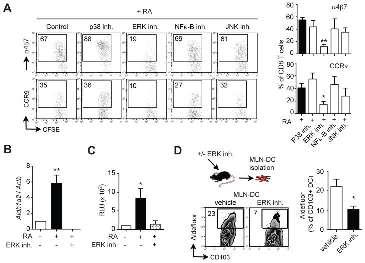
Background & aims: Gut-associated dendritic cells (DC) metabolize vitamin A into all-trans retinoic acid (RA), which is required to induce lymphocytes to localize to the gastrointestinal tract and promotes the differentiation of Foxp3+ regulatory T cells and IgA antibody-secreting cells. We investigated whether RA functions in a positive-feedback loop in DC to induce its own synthesis.

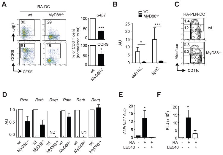
Methods: We measured levels of retinoids in intestinal tissues from mice and assessed the role of RA in the functional specialization of gut-associated DC in cell cultures and mice. We used pharmacologic antagonists to determine the signaling pathways involved in regulation of DC and used MyD88-/- mice to determine the contribution of Toll-like receptor signaling in RA-mediated effects on DC.

Results: The concentration of retinoids decreased in a proximal-to-distal gradient along the intestine, which correlated with the activity of gut-specific DC. Importantly, RA regulated the ability of gut-associated DC to produce RA, induce T cells to localize to the gastrointestinal tract, and generate regulatory T cells and IgA-secreting cells. RA was sufficient to induce its own production by extraintestinal DC in vitro and in vivo. RA-mediated regulation of DC required signaling through the mitogen-activated protein kinase signaling pathway and unexpectedly required MyD88, which is conventionally associated with Toll-like receptor, interleukin-1, and interleukin-18 signaling.

Conclusions: RA is necessary and sufficient to induce DC to regulate T-cell localization to the gastrointestinal tract and IgA secretion. Our findings also indicate crosstalk between the RA receptor and MyD88-dependent Toll-like receptor signaling pathways.


Conflict of interest statement

Conflicts of interest: The Authors disclose no conflicts

Figures

Figure 1
Figure 1. DC ability to imprint gut homing correlates with retinoid levels in the gut
(A) Quantification of retinyl esters, retinol and RA in duodenum (duo), jejunum (jej), ileum (ile) and colon (n=6). (B) LP-DC or PP-DC from duo, jej, ile, colon were used to activate naïve CD8 T cells. After 4–5 days T cells were analyzed for α4β7 and CCR9 expression (n=5). (C) DC were used to activate naïve CD4 T cells. After 4 days T cells were analyzed for Foxp3 expression. FACS plots are representative of two independent experiments. (D) Raldh activity in DC (n=4). (E) Luciferase activity in DC from DR5-luciferase mice (n=4). Mean ± SEM, *p<0.05, **p<0.01, ***p<0.001
Figure 2
Figure 2. RA is necessary in vivo for gut-associated DC education
(A–G) DC were isolated from mice on a vitamin A deficient (VAD) or control diet. (A) DC were used to activate naïve CD8 T cells. After 4–5 days T cells were analyzed for α4β7 and CCR9 expression (n=4). (B) DC were used to activate naïve CD4 T cells. After 4–5 days T cells were analyzed for Foxp3 expression. FACS plots are representative of two independent experiments. (C) DC were co-cultured with naïve B cells activated with anti-IgM plus IL-5. After 4 days the co-cultures were analyzed for intracellular IgA in B220Int cells and IgA in the supernatant (n=4). (D) Aldh1a2 mRNA and (E) Raldh activity in CD11c+ and CD11c+CD103+ DC (n=5). (F) Raldh activity in CD11c+ DC from control or VAD mice ± oral RA supplementation. FACS plots are representative of two independent experiments. Mean ± SEM, *p<0.05, **p<0.01
Figure 3
Figure 3. RA is sufficient to confer DC with RA synthesizing capacity in vitro
(A) Aldh1a2 mRNA in Spleen-DC incubated for 24 h with 100 nM all-trans RA (RA) or 100 nM of the indicated nuclear receptor agonists. HX630 and PA024 were used at 1 or 10 μM, with similar results (n=5–10). (B–F) Spleen-DC were incubated for 24 h with or without 100 nM RA (RA-DC and UT-DC, respectively) and analyzed for (B) Raldh activity (n=5) or (C) washed, pulsed with antigen and used to activate naïve CD8 T cells from TCR transgenic OT-1xRAG2−/− mice in FBS-free media ± 50 nM retinol. After 4–5 days T cells were analyzed for α4β7 and CCR9 expression (n=7). (D) Co-cultures were performed as described in (C) either in the presence or absence of the Raldh inhibitor DEAB (n=7). (E) Competitive homing experiment between CD8 T cells activated with RA-DC or UT-DC (n=5). (F) Naïve CD4 T cells from OT-1xRAG2−/− mice were activated with UT-DC or RA-DC. Five days later CD4 T cells were analyzed for Foxp3 expression (n=3).
Figure 4
Figure 4. RA is sufficient to confer DC with RA synthesizing capacity in vivo
(A) Wild type mice were supplemented orally with RA (400 μg/dose) every other day for 6 days. After that, MLN-DC and PLN-DC were isolated and analyzed for the expression of (B) Aldh1a2 mRNA and (C) Raldh activity (n=5). (D) PLN-DC from control or RA-treated mice were used to activate naïve CD8 T cells in FBS-free media ± retinol and either in the presence or absence of DEAB. FACS plots are representative of two independent experiments. Mean ± SEM, *p<0.05, **p<0.01, ***p<0.001
Figure 5
Figure 5. DC education requires ERK/MAPK signaling
(A–C) Spleen-DC were incubated for 24 h with or without 100 nM RA and in the presence or the absence of inhibitors for p38 (SB203580, 10 μM), ERK (U0126, 10 μM), NF-κB (SN50, 50 μM) or JNK (SP600125, 50 μM). (A) DC were used to activate naïve CD8 T cells. After 4–5 days T cells were analyzed for α4β7 and CCR9 expression (n=9). (B) Aldh1a2 mRNA expression in DC (n=3). (C) Spleen-DC from DR5-luciferase mice were incubated for 24 h with or without RA and in the presence or absence of the ERK inhibitor (U0126, 10 μM) and analyzed for their luciferase activity (n=3). (D) Wild type mice were orally treated with the ERK inhibitor PD0325901 (25 μg/g/dose) every other day for 6 days. After that, CD11c+ MLN-DC were analyzed for CD103 expression and Raldh activity (n=5). Mean ± SEM, *p<0.05, **p<0.01
Figure 6
Figure 6. MyD88 is required for RA-mediated DC education
(A–B) Wild type or MyD88−/− Spleen-DC were incubated with 100 nM RA for 24 h. (A) DC were used to activate naïve CD8 T cells. After 4–5 days T cells were analyzed for α4β7 and CCR9 expression (n=5). (B) Aldh1a2 (n=4) or Tgm2 (n=5) mRNA expression in DC. (C) Wild type or MyD88−/− mice were supplemented orally with RA (400 μg/dose) every other day for 6 days and PLN-DC were analyzed for Raldh activity (n=7). (D) Spleen-DC from wild type or MyD88−/− mice were analyzed for their expression of Rxra, Rxrb. Rxrg, Rara, Rarb and Rarg mRNA (n=4–6). ND: not detected. (E) Spleen-DC were incubated for 24 h with or without 100 nM RA and either in the presence or absence of the RAR® inhibitor LE540 (1 μM) and then analyzed for Aldh1a2 mRNA expression (n=3). (F) Spleen-DC from DR5-luciferase mice were incubated for 24 h with or without 100 nM RA and either in the presence or absence of LE540 and then analyzed for their luciferase activity (n=3). Mean ± SEM, *p<0.05, ***p<0.001
Figure 7
Figure 7. TLR signals contribute to RA-mediated human and murine DC education
(A) Monocyte-derived DC (Mo-DC) from healthy donors were treated with either RA (100 nM), Pam3CSK4 (0.5 μg/ml) or both from day 3 of differentiation. Raldh activity was analyzed at day 6 in CD11c+ cells (n=4). FACS plots show representative results in Mo-DC from two donors. (B) Mo-DC pretreated with either RA, Pam3CSK4 or both were washed and cultured with allogenic CD4 T cells activated with anti-CD3 plus anti-CD28 and hTGF-β1 (2 ng/ml). After 4 days, CD4 T cells were analyzed for Foxp3 expression. FACS plots show one representative experiment out of two. (C) Spleen-DC were incubated for 24 h in the presence of 100 nM of RA, Pam3CSK4 (0.5 μg/ml) or both, washed and used to activate naive CD8 T cells. After 4 days, T cells were analyzed for their expression of α4β7 and CCR9. FACS plots are representative of three experiments. (D) Human Mo-DC were treated at day 6 with or without 100 nM of RA, Pam3CSK4 (0.5 μg/ml) or both for 24 h, washed and co-cultured with total human T cells activated with plate-bound anti-CD3 plus anti-CD28 antibodies. After 6 days, CD4 and CD8 T cells were analyzed for their expression of α4β7 (n=3). (E) Spleen-DC were incubated for 24 h in the presence of 100 nM of RA, Pam3CSK4 (0.5 μg/ml) or both, washed and co-cultured with naïve B cells activated with anti-IgM plus IL-5. After 4 days IgA levels were measured in the culture supernatants (n=7). Mean ± SEM, *p<0.05, ***p<0.001

References

    1. Mora JR. Homing imprinting and immunomodulation in the gut: role of dendritic cells and retinoids. Inflamm Bowel Dis. 2008;14:275–89. - PubMed
    1. Eksteen B, Adams DH. GSK-1605786, a selective small-molecule antagonist of the CCR9 chemokine receptor for the treatment of Crohn’s disease. IDrugs. 2010;13:472–781. - PubMed
    1. Mora JR, Bono MR, Manjunath N, Weninger W, Cavanagh LL, Rosemblatt M, von Andrian UH. Selective imprinting of gut-homing T cells by Peyer’s patch dendritic cells. Nature. 2003;424:88–93. - PubMed
    1. Johansson-Lindbom B, Svensson M, Wurbel MA, Malissen B, Marquez G, Agace W. Selective generation of gut tropic T cells in gut-associated lymphoid tissue (GALT): requirement for GALT dendritic cells and adjuvant. J Exp Med. 2003;198:963–969. - PMC - PubMed
    1. Mora JR, Iwata M, Eksteen B, Song SY, Junt T, Senman B, Otipoby KL, Yokota A, Takeuchi H, Ricciardi-Castagnoli P, Rajewsky K, Adams DH, von Andrian UH. Generation of gut-homing IgA-secreting B cells by intestinal dendritic cells. Science. 2006;314:1157–60. - PubMed

Publication types

MeSH terms

Substances