. 2011 May 20;42(4):489-99.
doi: 10.1016/j.molcel.2011.04.015.

Poly(ADP-ribose) regulates stress responses and microRNA activity in the cytoplasm

Affiliations

Poly(ADP-ribose) regulates stress responses and microRNA activity in the cytoplasm

Anthony K L Leung et al. Mol Cell. .

Abstract

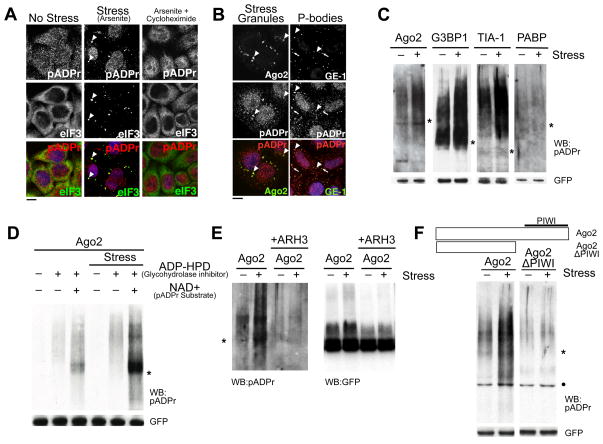
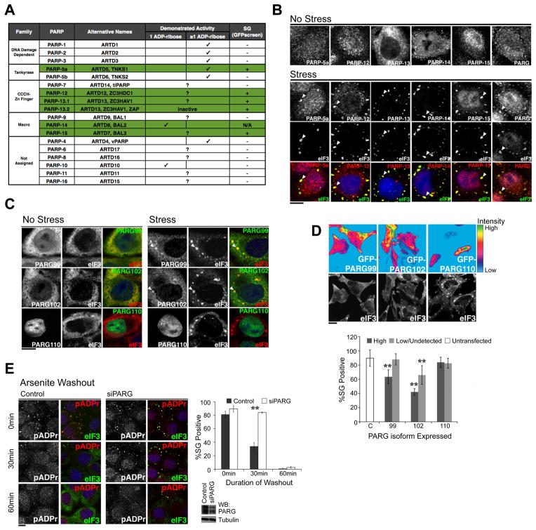
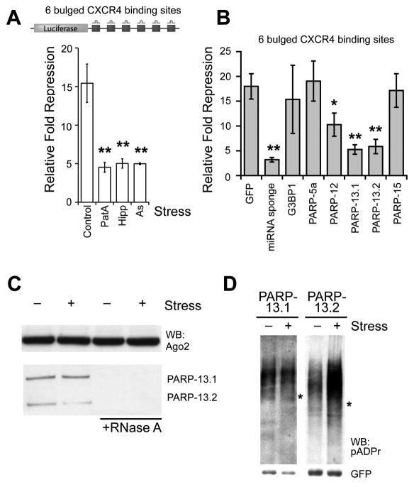
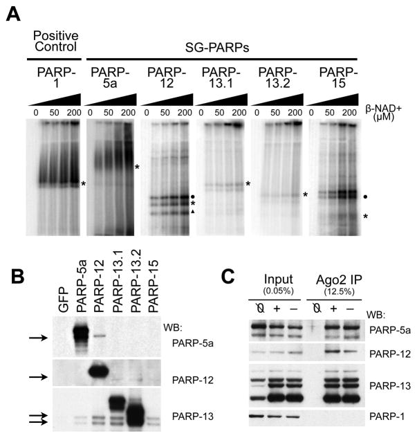
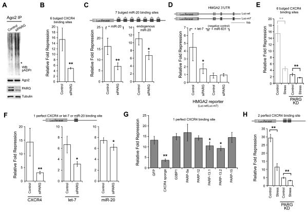
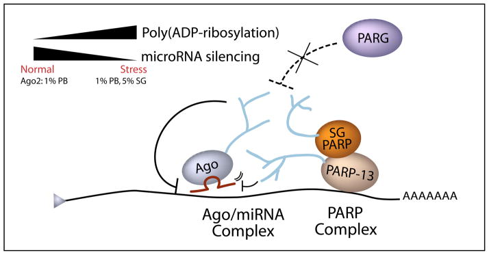
Poly(ADP-ribose) is a major regulatory macromolecule in the nucleus, where it regulates transcription, chromosome structure, and DNA damage repair. Functions in the interphase cytoplasm are less understood. Here, we identify a requirement for poly(ADP-ribose) in the assembly of cytoplasmic stress granules, which accumulate RNA-binding proteins that regulate the translation and stability of mRNAs upon stress. We show that poly(ADP-ribose), six specific poly(ADP-ribose) polymerases, and two poly(ADP-ribose) glycohydrolase isoforms are stress granule components. A subset of stress granule proteins, including microRNA-binding Argonaute family members Ago1-4, are modified by poly(ADP-ribose), and such modification increases upon stress, a condition when both microRNA-mediated translational repression and microRNA-directed mRNA cleavage are relieved. Similar relief of repression is also observed upon overexpression of specific poly(ADP-ribose) polymerases or, conversely, upon knockdown of glycohydrolase. We conclude that poly(ADP-ribose) is a key regulator of posttranscriptional gene expression in the cytoplasm.


Figures

Figure 1
Figure 1. pADPr is enriched in SGs upon multiple types of stresses and modifies specific cytoplasmic RNA-binding proteins dependent on RNA-binding domain
(A) pADPr staining using LP96-10 antibodies in HeLa cells untreated, or treated with 100 μM arsenite for 60 min, or for 30 min followed by 100 μM arsenite +100 μg/ml cycloheximide for 30 min. Arrowheads, SGs; scale bar, 10 μm. (B) HeLa cells treated with 100 μM arsenite were stained for pADPr, SG component Ago2 (arrowheads) or PB component GE-1 (arrows). DNA was stained with Hoeschst 33342 (blue); scale bar, 10 μm. (C) Immunoprecipitates of 4 GFP-tagged SG-localized RNA-binding proteins from cells treated with or without 20 nM pateamine A were probed for pADPr. (D) Immunoprecipitates of GFP-Ago2 from cells treated with or without 20 nM pateamine A were probed for pADPr. The cell extracts either included or excluded 1 μM ADP-HPD, and with or without 1 mM NAD+ before immunoprecipitation by anti-GFP. (E) pADPr modification of Ago2 from cells treated with or without 20 nM pateamine A was verified by treating the immunoprecipitates with ARH3. The immunoprecipiates were probed for pADPr (left) and GFP (right). (F) Immunoprecipitates of wild-type and PIWI mutant of GFP-Ago2 from cells treated with or without 20 nM pateamine A were probed for pADPr. For panels C-F, cell extracts included 1μM ADP-HPD unless stated otherwise; shown are western blots for pADPr (LP96-10) and GFP levels in each immunoprecipitate. Asterisks indicate the position of the corresponding GFP-tagged RNA-binding protein constructs. Black dots indicate non-specific binding to BSA by LP96-10. See also Figure S1.
Figure 2
Figure 2. Specific PARPs and PARG isoforms localize in the cytoplasmic SGs and the level of PARG modulates the kinetics of SG assembly and disassembly
(A) Summary of SG localization screen of PARP family. Green shading indicates SG-PARPs as determined by GFP-PARP fusions or PARP specific antibodies. (B) HeLa cells were treated with or without 100 μM arsenite for 60 min and stained using antibodies against SG-PARPs and PARG. (C) HeLa cells expressing GFP-tagged PARG isoforms were treated with or without 250 μM arsenite for 30 min. (D) Overexpression of cytoplasmic PARG isoforms inhibits SG assembly. Experiment performed as in panel C, but heat map shows level of GFP-PARG isoforms (99, 102, 110) compared with untransfected control (C). Accompanying graph shows quantitation of image data; ≥200 cells for each condition from at least six independent fields. Cells with GFP intensity above background are classified as ‘High’ while cells with intensity indistinguishable from background levels as ‘Low/Undetected’. Paired t-test p < 0.01 (**), derived from comparison to untransfected control; error bars indicates SD. (E) pADPr hydrolysis is required for SG disassembly. Shown are representative images taken 0, 30, and 60 min after washout of 30 min 100 μM arsenite treatment in control and PARG knockdown HeLa cells. Quantitation: ≥100 cells for each condition, n = 3. Paired t-test p < 0.01 (**) and error bars indicate SD. Accompanying blot shows level of PARG knockdown with tubulin as loading control. For panels B-E, pADPr were stained by LP96-10 antibodies, SGs (arrowheads) by anti-eIF3 and DNA by Hoechst 33342 (blue); scale bars = 10 μm. See also Figure S2 and Movie S1.
Figure 3
Figure 3. Stress or PARP-13 overexpression alleviates miRNA-mediated repression
(A) miRNA activity assay in untreated 293T cells or cells treated with 30 nM pateamine A (PatA), 1 μM hippuristanol (Hipp) or 250 μM arsenite (As) for 2 hr, where relative fold repression was measured as the activity of luciferase (upper panel) in the presence of the targeting siRNA normalized to a control siRNA; n = 3. (B) miRNA activity assay upon overexpression of SG-PARPs. Relative fold repression was measured as in panel A; n = 4. For panels A and B, error bars indicate SD; paired t-test p < 0.05 (*) and < 0.01 (**). (C) Antibodies against endogenous Ago2 was used for immunoprecipitation from cytoplasmic extract of HeLa cells treated with or without 250 μM arsenite for 30 min. On the right, the extract was pre-treated with 200 μg/ml RNase A for 20 min at 25°C. (D) Immunoprecipitates of GFP-tagged PARP-13.1 or -13.2 from cells treated with or without 20 nM pateamine A for 30 min were probed for pADPr, where cell extracts included 1 μM ADP-HPD. Asterisks indicate the position of the corresponding GFP-tagged PARPs. See also Figure S3.
Figure 4
Figure 4. PARP-13 family members are poly(ADP-ribosylated) by other SG-PARPs
(A) in vitro ADP-ribosylating assay for PARP-1, -5a, -12, -13.1, -13.2 and -15. HeLa S3 cells were transfected with individual GFP-tagged SG-PARPs and GFP-PARP immunoprecipitates were washed twice with 450 mM NaCl then once with 150 mM NaCl. The immunoprecipitates were incubated with 0, 25, 50, 100 or 200 μM NAD+ (with 1/175 fold of P32-labeled NAD+) at 16°C for 30 min, separated on a 6% SDS-PAGE gel and visualized by autoradiography. Asterisks indicate the position of the corresponding GFP-tagged SG-PARPs; circles and triangle indicate the endogenous position of PARP-13.1 and PARP-13.2 respectively. (B) Western blots of the immunoprecipitates from panel A were probed with PARP-5a, -12 and -13 (antibodies for PARP-15 are not good for detecting endogenous protein). Immunoprecipitates from cells transfected with GFP were used as a negative control. (C) HeLa S3 cells were transfected with GFP-tagged Ago2 and cells either treated with (+) or without (−) 20 nM pateamine A for 30 min. Untransfected cells treated with 20 nM pateamine A were used as a negative control (Ø). The cytoplasmic lysates were immunoprecipitated with anti-GFP and washed thrice with cytoplasmic lysis buffer. The input and immunoprecipitates were probed with antibodies against PARP-1, -5a, -12 and -13. See also Figure S4.
Figure 5
Figure 5. PARG knockdown alleviates miRNA-mediated repression and miRNA-directed cleavage
(A) pADPr modification levels of endogenous Ago2 in HeLa S3 cells transfected with 25 nM control siRNA or siPARG for 48 hr. Asterisk indicates where Ago2 migrated. Shown are western blots for Ago2, PARG and tubulin. (B) 293T cells were transfected with 25 nM control siRNA or siPARG for 72 hr. Relative fold repression was measured as in Figure 3A; n = 3. (C) PARG knockdown effect observed in luciferase reporter with 7 artificial miR-20 binding sites. The relative fold repression was calculated by the amount of expression of the construct normalized to a construct with all binding sites mutated at their seed positions. The assay was tested with exogenous addition of miR-20 (left) or with endogenous miR-20 (right); n = 4. (D) PARG knockdown effect observed in luciferase reporter with endogenous HMGA2 3′UTR. The relative fold repression is calculated by the amount of expression by the wild-type construct (Luc-wt) normalized to the mutant construct Luc-m7; n = 4. (E) siPARG-transfected cells were either treated with or without 30 nM pateamine A for 2 hr (right). As a comparison, part of Figure 3A is reproduced here on the left to show cells transfected with a control siRNA. (F) The effect of PARG knockdown on miRNA-directed cleavage was examined for luciferase construct with 1 perfect siCXCR4, let-7 or miR-20 binding site; n = 4 in each case. (G) The effect of SG-PARP overexpression on miRNA-directed cleavage assay as in panel F; n = 5. (H) The effect of stress on miRNA-directed cleavage was tested with a luciferase reporter with 2 perfect binding sites for siCXCR4 using the same transfection conditions and drug treatment as in Panel E; n = 3. For panels B-H, error bars indicate SD; paired t-test p < 0.05 (*) and < 0.01 (**). See also Figure S5.
Figure 6
Figure 6. A working model: A high local concentration of pADPr at miRNA complex results in relief of miRNA silencing
Upon stress, multiple proteins including all Argonaute family members and PARP-13.1/2 complex are increasingly modified by pADPr. Such increase in poly(ADP-ribosylation) during stress could be due to increase in PARP activity and/or decrease in PARG activity (dotted line). High concentration of pADPr near the Argonaute/miRNA complex might disrupt electrostatic interaction or cause steric hindrance for effective miRNA silencing. Similar relief of miRNA silencing is also observed upon overexpression of PARP-13 or, conversely, upon knockdown of PARG.

References

    1. Ahel D, Horejsi Z, Wiechens N, Polo SE, Garcia-Wilson E, Ahel I, Flynn H, Skehel M, West SC, Jackson SP, et al. Poly(ADP-ribose)-dependent regulation of DNA repair by the chromatin remodeling enzyme ALC1. Science. 2009;325:1240–1243. - PMC - PubMed
    1. Anderson P, Kedersha NL. Stress granules: the Tao of RNA triage. Trends Biochem Sci. 2008;33:141–150. - PubMed
    1. Bartel DP. MicroRNAs: target recognition and regulatory functions. Cell. 2009;136:215–233. - PMC - PubMed
    1. Bhattacharyya S, Habermacher R, Martine U, Closs E, Filipowicz W. Relief of microRNA-Mediated Translational Repression in Human Cells Subjected to Stress. Cell. 2006;125:1111–1124. - PubMed
    1. Chang P, Coughlin M, Mitchison TJ. Interaction between Poly(ADP-ribose) and NuMA contributes to mitotic spindle pole assembly. Mol Biol Cell. 2009;20:4575–4585. - PMC - PubMed

Publication types

MeSH terms