

Reduction of dorsolateral prefrontal cortex gray matter in late-life depression
- PMID: 21596532
- PMCID: PMC3105213
- DOI: 10.1016/j.pscychresns.2011.01.003
Reduction of dorsolateral prefrontal cortex gray matter in late-life depression
Abstract
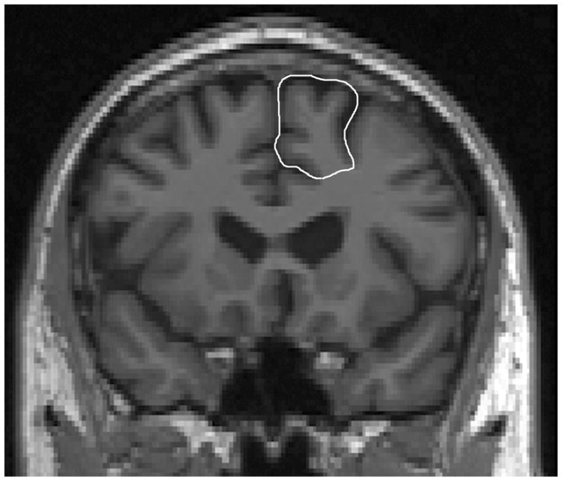
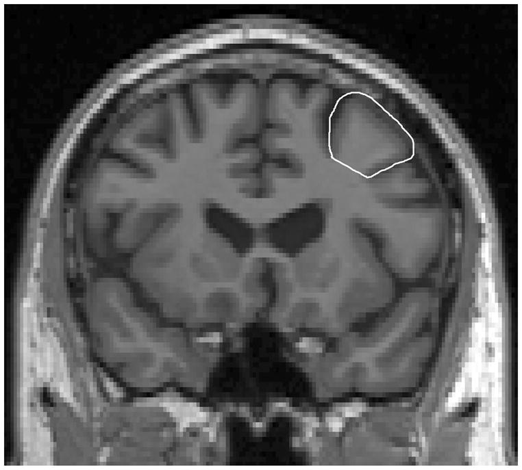
Postmortem studies have documented abnormalities in the dorsolateral prefrontal cortex (dlPFC) in depressed subjects. In this study we used magnetic resonance imaging to test for dlPFC volume differences between older depressed and non-depressed individuals. Eighty-eight subjects meeting DSM IV criteria for major depressive disorder and thirty-five control subjects completed clinical evaluations and cranial 3T magnetic resonance imaging. After tissue types were identified using an automated segmentation process, the dlPFC was measured in both hemispheres using manual delineation based on anatomical landmarks. Depressed subjects had significantly lower gray matter in the left and right dorsolateral prefrontal cortex (standardized to cerebral parenchyma) after controlling for age and sex. Our study confirmed the reduction of dorsolateral prefrontal cortex in elderly depressed subjects, especially in the gray matter. These regional abnormalities may be associated with psychopathological changes in late-life depression.
Copyright © 2011 Elsevier Ireland Ltd. All rights reserved.
Figures
References
-
- Alexopoulos GS, Meyers BS, Young RC, Kakuma T, Silbersweig D, Charlson M. Clinically defined vascular depression. American Journal of Psychiatry. 1997;154(4):562–565. - PubMed
-
- Bench CJ, Friston KJ, Brown RG, Frackowiak RS, Dolan RJ. Regional cerebral blood flow in depression measured by positron emission tomography: the relationship with clinical dimensions. Psychological Medicine. 1993;23(3):579–590. - PubMed
-
- Bench CJ, Friston KJ, Brown RG, Scott LC, Frackowiak RS, Dolan RJ. The anatomy of melancholia--focal abnormalities of cerebral blood flow in major depression. Psychological Medicine. 1992;22(3):607–615. - PubMed
-
- Biver F, Goldman S, Delvenne V, Luxen A, De Maertelaer V, Hubain P, Mendlewicz J, Lotstra F. Frontal and parietal metabolic disturbances in unipolar depression. Biological Psychiatry. 1994;36(6):381–388. - PubMed
-
- Braver TS, Cohen JD, Nystrom LE, Jonides J, Smith EE, Noll DC. A parametric study of prefrontal cortex involvement in human working memory. Neuroimage. 1997;5(1):49–62. - PubMed
Publication types
MeSH terms
Grants and funding
LinkOut - more resources
Full Text Sources
Other Literature Sources
Medical
