. 2011 Jun 7;21(11):948-54.
doi: 10.1016/j.cub.2011.04.025. Epub 2011 May 19.

The Arf GAP CNT-2 regulates the apoptotic fate in C. elegans asymmetric neuroblast divisions

Affiliations

The Arf GAP CNT-2 regulates the apoptotic fate in C. elegans asymmetric neuroblast divisions

Aakanksha Singhvi et al. Curr Biol. .

Abstract

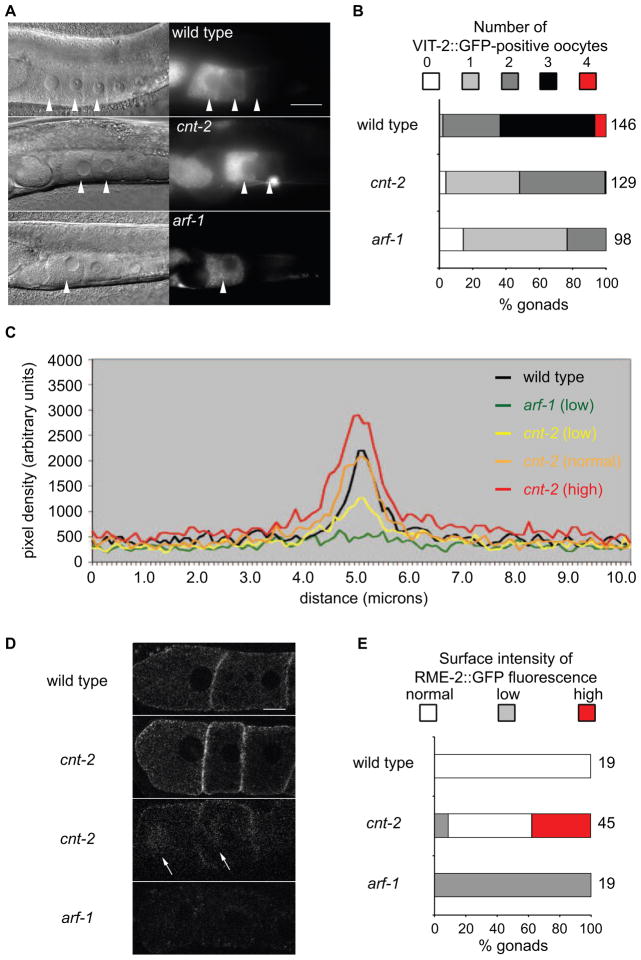
During development, all cells make the decision to live or die. Although the molecular mechanisms that execute the apoptotic program are well defined, less is known about how cells decide whether to live or die. In C. elegans, this decision is linked to how cells divide asymmetrically [1, 2]. Several classes of molecules are known to regulate asymmetric cell divisions in metazoans, yet these molecules do not appear to control C. elegans divisions that produce apoptotic cells [3]. We identified CNT-2, an Arf GTPase-activating protein (GAP) of the AGAP family, as a novel regulator of this type of neuroblast division. Loss of CNT-2 alters daughter cell size and causes the apoptotic cell to adopt the fate of its sister cell, resulting in extra neurons. CNT-2's Arf GAP activity is essential for its function in these divisions. The N terminus of CNT-2, which contains a GTPase-like domain that defines the AGAP class of Arf GAPs, negatively regulates CNT-2's function. We provide evidence that CNT-2 regulates receptor-mediated endocytosis and consider the implications of its role in asymmetric cell divisions.


Figures

Figure 1
Figure 1
cnt-2 mutations disrupt the asymmetric divisions of the Q.p neuroblast. (A) The Q.p lineage. (B) The Q.p lineage in cell fate and cell death mutant animals. (C) Frequency of extra A/PVM neurons in cnt-2 mutants. All of the animalss carried the Pmec-4::gfp transgene zdIs5. cnt-2 mutant lineages either produced a single A/PVM or two A/PVMs (% Q.p lineages with extra A/PVMs). Animals were mutant for both maternal and zygotic cnt-2 (m−z−) or only for zygotic cnt-2 (m+z−). For this and subsequent graphs, the number of Q.p lineages scored is above each genotype. (D, E) Alleles analyzed were cnt-2(gm377), ced-4(n1162) and arf-1(ok796). Animals contained the Pegl-17::gfp transgene ayIs9. (D) Ratio of the Q.p daughter cell sizes. Above the bars are representative photomicrographs and drawings of Q.p daughter cells. (E) Fluorescence photomicrographs from time-lapse confocal recordings of wild-type, cnt-2 and arf-1 Q.p divisions. Also see supplemental movies. Numbers at the bottom left corner of each frame represents the time in seconds. Arrows indicate the position of the cleavage furrow. Normally, the QL.a cell migrates over the top of Q.p towards the tail and then divides. QL.ap, the PQR neuron, continues to migrate toward the tail, but QL.aa dies. The arrowhead indicates a QL.aa cell that did not die. Scale bar represents 2 m.
Figure 2
Figure 2
CNT-2 and the role of its GAP domain in the Q.p division. (A) Structure of CNT-2B. GLD: GTPase-Like Domain; PH and DM: split Pleckstrin Homolog Domain; GAP: Arf GTPase Activating Protein domain; A: Ankyrin repeats. (B) Structure of the A, B and C mRNAs. Boxes represent exons, lines introns. Shaded boxes represent coding sequences; the open box represents the 5′ UTR of isoform A. The slashes in intron 1 of the B isoform indicate that intron length is larger than drawn. The tm2328 and gm377 deletions are predicted to shift the reading frame. (C and D) Effects of cnt-2 transgenes on the Q.p lineage. See Figure 1C for presentation. The mutation that changes a conserved arginine to lysine at position 709 (CNT-2B(RK)::GFP) abrogated the GAP activity of AGAP1 while still allowing the cognate Arf to bind [9]. The cysteine to serine changes at positions 681 and 684 within the zinc-finger motif (CNT-2-2B(CCSS)::GFP) are predicted to render the Arf GAP incapable of binding its cognate Arf [9, 10]. We generated one transgenic line for cnt-2B, five for cnt-2B::gfp, two each for the mutated versions of cnt-2B::gfp and ΔNcnt-2B::gfp, and three for ΔNcnt-2B::gfp. Each of the lines for a particular construct was tested and gave similar results. Data for only one line of each type is presented. GFP levels were similar for all of the transgenes. * P < 0.0001 (two-sample proportion test).
Figure 3
Figure 3
Cell autonomous roles and genetic interactions for arf-1 and cnt-2. Alleles used were arf-1(ok796), arf-6(tm1447), ced-3(n717), ced-4(n1162) and cnt-2(gm377). (A) Ratio of the Q.p daughter cell sizes. See Figure 1D for presentation of results. (B, C) See Figure 1C for presentation of results. (B) Expression of a cnt-2 or an arf-1 cDNA from the mab-5 promoter rescued the QL (PVM) but not the QR (AVM) defects of cnt-2 or arf-1; ced-3 mutant animals, respectively. The frequency of extra AVMs (open bars) and PVMs (solid bars) are presented separately. (C) The roles of arf-1 and arf-6 in the Q.p division. (A, C) Because arf-1 and cnt-2 are linked on LG III, unc-32(e189) and dpy-18(e364) are visible markers used to construct the arf-1 cnt-2 double mutant. ^The arf-1 cnt-2 chromosome also contained an unc-32 mutation. #The cnt-2 control in C contained the unc-32 and dpy-18 mutations. * P < 0.0001, ** P <0.3, ***P <0.01.
Figure 4
Figure 4
CNT-2 regulates receptor-mediated endocytosis. arf-1(ok796) and cnt-2(gm377) were used. (A) Nomarski (left panels) and fluorescence photomicrographs (right panels) of wild-type, cnt-2, and arf-1 animals containing [vit-2::gfp]. Arrowheads indicate the positions of individual oocytes. The scale bar represents 30 microns. (B) Quantification of the number of VIT-2::GFP-positive oocytes in a gonad arm. To the right of the bars are the number of gonad arms scored. P < 0.0001 for both arf-1 and cnt-2 compared to wild-type using a two-sample test for proportions. (C) Plots of line scans through the first two oocytes of wild-type, arf-1 and cnt-2 mutants that contain the rme-2::gfp transgene. ImageJ was used to draw a line perpendicular to, and centered on, the boundary between the first and the second oocyte on 12bit depth confocal images. The plot profile for this line was recorded, and five independent profiles were averaged for each gonad. Based on the maximum intensity of fluorescence (Imax) measured in arbitrary units (AU), gonads were grouped into low (Imax<1000 AU), normal (1000AU<Imax<2500AU) or high (Imax>2500) fluorescence classes. The three classes of RME-2::GFP surface intensity quantified in E are depicted here. The range of the normal class is illustrated by the wild-type and cnt-2 scans. (D) Confocal images RME-2::GFP in wild-type and mutant oocytes. Arrows indicate juxtanuclear dots. The scale bar represents 10 microns. The number of oocytes scored is to the right of the bar. (E) Quantification of RME-2::GFP levels at the oocyte cell surface. P < 0.0001 for both arf-1 and cnt-2 compared to wild-type using a two-sample test for proportions. Figure S2 describes the effects of interactions between arf-1 and cnt-2 on RME-2::GFP distribution.

References

    1. Sulston JE, Horvitz HR. Post-embryonic cell lineages of the nematode, Caenorhabditis elegans. Dev Biol. 1977;56:110–156. - PubMed
    1. Sulston JE, Schierenberg E, White JG, Thomson JN. The embryonic cell lineage of the nematode Caenorhabditis elegans. Dev Biol. 1983;100:64–119. - PubMed
    1. Singhvi A, Garriga G. Asymmetric divisions, aggresomes and apoptosis. Trends Cell Biol. 2009;19:1–7. - PMC - PubMed
    1. Cordes S, Frank CA, Garriga G. The C. elegans MELK ortholog PIG-1 regulates cell size asymmetry and daughter cell fate in asymmetric neuroblast divisions. Development. 2006;133:2747–2756. - PubMed
    1. Ellis HM, Horvitz HR. Genetic control of programmed cell death in the nematode C. elegans. Cell. 1986;44:817–829. - PubMed

Publication types

MeSH terms

Substances

LinkOut - more resources