Review
. 2011 Aug;36(8):397-404.
doi: 10.1016/j.tibs.2011.04.003. Epub 2011 May 17.

Degrade, move, regroup: signaling control of splicing proteins

Affiliations
Review

Degrade, move, regroup: signaling control of splicing proteins

Florian Heyd et al. Trends Biochem Sci. 2011 Aug.

Abstract

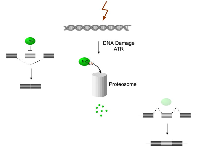
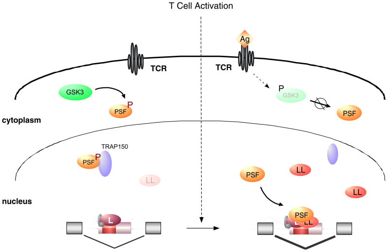
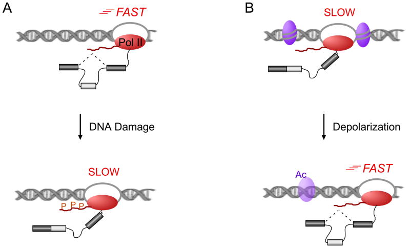
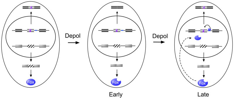
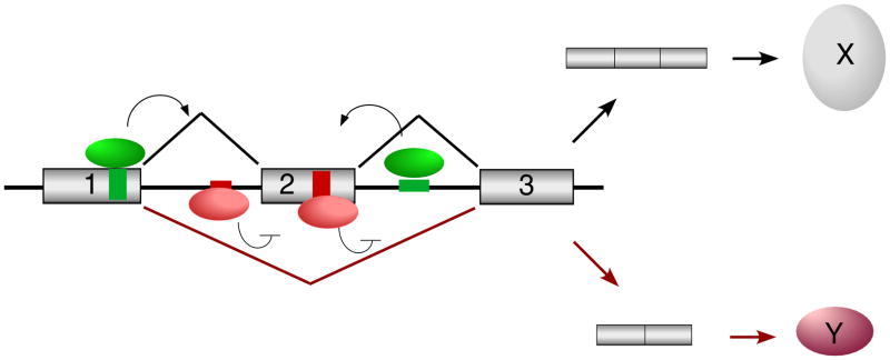
With recent advances in microarrays and sequencing it is now relatively straightforward to compare pre-mRNA splicing patterns in different cellular conditions on a genome-wide scale. Such studies have revealed extensive changes in cellular splicing programs in response to stimuli such as neuronal depolarization, DNA damage, immune signaling and cellular metabolic changes. However, for many years our understanding of the signaling pathways responsible for such splicing changes was greatly lacking. Excitingly, over the past few years this gap has begun to close. Recent studies now suggest notable trends in the mechanisms that link cellular stimuli to downstream alternative splicing events. These include regulated synthesis or degradation of splicing factors, differential protein-protein interactions, altered nuclear translocation and changes in transcription elongation.


Figures

Fig.1
Fig.1. Regulated protein turnover as a means to control alternative splicing
An example of how the regulated degradation of RNA binding proteins can modulate alternative splicing. Tra2 levels are reduced by proteasomal degradation in response to ataxia telangiectasia and Rad3 related (ATR) signaling, most likely through ubiquitin (Ub)-mediated degradation. The reduction in Tra2 protein alleviates normal Tra2 repression of Taf1 alternative exons (light grey boxes). Reproduced, with permission from [23].
Fig. 2
Fig. 2. Altered protein–protein interactions and induced protein expression regulate CD45 alternative splicing during T cell activation
(i) In resting T cells, the RNA binding protein PSF is phosphorylated (P) by GSK3 and forms a tight complex with TRAP150. In this complex, PSF is unable to bind CD45 RNA and therefore does not participate in splicing regulation. (ii) Upon T cell activation by antigen (Ag) binding to the T cell receptor (TCR), GSK3 activity decreases due to an inhibiting phosphorylation. This leads to accumulation of newly translated, hypophosphorylated PSF which is not bound to TRAP150 and thus is recruited to the CD45 exon silencer to increase exon exclusion in activated T cells. HnRNP LL also contributes to increased CD45 exon exclusion in activated T cells. hnRNP LL mRNA and protein levels increase upon T cell activation, thus providing an example of a splicing regulator that is controlled by signal-induced changes in transcription.
Fig. 3
Fig. 3. Signal-induced changes in Pol II processivity regulate cotranscriptional splicing
In the kinetic coupling model, alternative splicing can be regulated by the speed of RNA polymerase II (Pol II; red oval) such that a slower polymerase leaves the spliceosome more time to recognize exons with suboptimal splice sites (light grey box), thus increasing inclusion. A) A model for DNA damage-induced alternative splicing of several apoptotic regulators, showed that phosphorylation (P) within the C-terminal domain (CTD, red wavy line) of Pol II leads to reduced Pol II speed and changed patterns of alternative splicing. B) Another mechanism to change Pol II processivity has been seen in neurons. Depolarization leads to loosening of the chromatin structure by changing the acetylation (Ac) of specific histones (purple ovals). This allows for increased Pol II elongation rates and leads to a consistent change in NCAM1 alternative splicing.
Fig. 4
Fig. 4. A feedback loop in signal-induced alternative splicing
CaRREs (purple box) and binding sites for FOX1 (blue line) represent two splicing regulatory elements known to regulate neural-specific genes. Upon neural depolarization (Depol), the CaRRE-responsive exons and exon 19 of FOX1 (striped box) are initially repressed. The skipping of FOX1 exon 19 results in expression of a form of FOX1 that is localized to the nucleus. For exons that are controlled by both CaRRE and FOX1 elements, this increase in nuclear FOX1 counters the activity of the CaRRE, thereby restoring exon inclusion.
Figure I
Figure I
Reciprocal regulation of alternative splicing by activator and repressor proteins

References

    1. Pan Q, et al. Deep surveying of alternative splicing complexity in the human transcriptome by high-throughput sequencing. Nature genetics. 2008;40:1413–1415. - PubMed
    1. Wang ET, et al. Alternative isoform regulation in human tissue transcriptomes. Nature. 2008;456:470–476. - PMC - PubMed
    1. Nilsen TW, Graveley BR. Expansion of the eukaryotic proteome by alternative splicing. Nature. 2010;463:457–463. - PMC - PubMed
    1. Lynch KW. Regulation of alternative splicing by signal transduction pathways. Adv Exp Med Biol. 2007;623:161–174. - PubMed
    1. Shin C, Manley JL. Cell signalling and the control of pre-mRNA splicing. Nature reviews. 2004;5:727–738. - PubMed

MeSH terms

LinkOut - more resources