. 2011 Jun 10;42(5):689-99.
doi: 10.1016/j.molcel.2011.04.020. Epub 2011 May 19.

A SIRT1-LSD1 corepressor complex regulates Notch target gene expression and development

Affiliations

A SIRT1-LSD1 corepressor complex regulates Notch target gene expression and development

Peter Mulligan et al. Mol Cell. .

Abstract

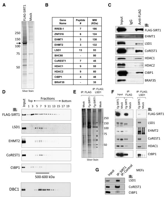
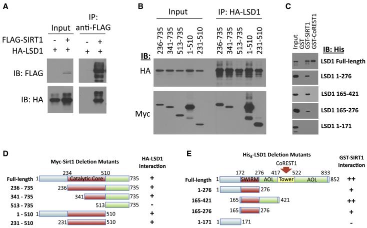
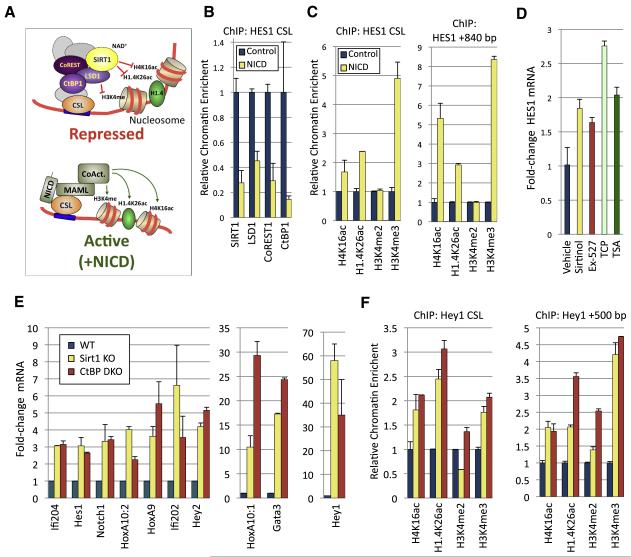
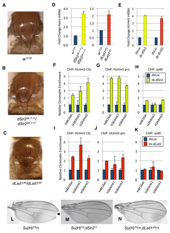
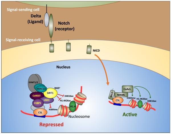
Epigenetic regulation of gene expression by histone-modifying corepressor complexes is central to normal animal development. The NAD(+)-dependent deacetylase and gene repressor SIRT1 removes histone H4K16 acetylation marks and facilitates heterochromatin formation. However, the mechanistic contribution of SIRT1 to epigenetic regulation at euchromatic loci and whether it acts in concert with other chromatin-modifying activities to control developmental gene expression programs remain unclear. We describe here a SIRT1 corepressor complex containing the histone H3K4 demethylase LSD1/KDM1A and several other LSD1-associated proteins. SIRT1 and LSD1 interact directly and play conserved and concerted roles in H4K16 deacetylation and H3K4 demethylation to repress genes regulated by the Notch signaling pathway. Mutations in Drosophila SIRT1 and LSD1 orthologs result in similar developmental phenotypes and genetically interact with the Notch pathway in Drosophila. These findings offer new insights into conserved mechanisms of epigenetic gene repression and regulation of development by SIRT1 in metazoans.


Figures

Figure 1
Figure 1. Identification and characterization of the SIRT1-LSD1-CtBP1 complex
(A) FLAG-tagged SIRT1 was transiently induced in a Tet-ON HEK293T stable cell line and nuclear extracts prepared. FLAG-SIRT1 and associated proteins were immunoaffinity-purified and eluted with FLAG peptide. Silver-stained SDS-PAGE gels showed that multiple polypeptides specifically associated with FLAG-SIRT1 as compared to control extracts from un-transfected cells. (B) Tandem mass spectrometry (MS-MS) identified numerous interacting proteins, including multiple components of the LSD1/CtBP1/CoREST1 complexes. (C) Immunoblotting was used to confirm the presence of core LSD1 complex components in FLAG-SIRT1 immunopurified material. (D) Analysis of FLAG-SIRT1 purified material by glycerol gradient sedimentation revealed co-sedimentation of LSD1, EHMT2, CoREST1, and CtBP1 (fractions 5 – 9, corresponding to M.W. markers 500-600 kDa). By contrast, another FLAG-SIRT1-interacting protein, DBC1, peaked in fractions 7 – 13. (E) FLAG-SIRT1 was purified as in (A), eluted under native conditions, followed by a second immunopurification using anti-LSD1 antibodies. Sequentially immunopurified proteins were eluted and analysed by SDS-PAGE/silver staining. (F) Immunoblotting analysis of sequentially-purified material from (E). (G) Immunoblot showing co-immunoprecipitation of endogenous LSD1, CoREST1 and CtBP1 with anti-SIRT1 in MEF nuclear extracts. Experiments in panels D – G were repeated three times with similar results.
Figure 2
Figure 2. Direct interaction between SIRT1 and LSD1
(A) Full-length FLAG-SIRT1 and HA-LSD1 co-immunopurify from transfected HEK293T cell nuclear extracts. Shown is an immunoblot analysis of FLAG-SIRT1 IP. (B) The indicated Myc-mSirt1 deletion mutants (D) were examined for co-immunopurification with HA-LSD1 from co-transfected HEK293T cell nuclear extracts. SIRT1 catalytic core region alone (231 – 510) was sufficient, for LSD1 binding. (C) Purified recombinant GST-SIRT1 interacted directly with purified recombinant His6-LSD1. GST-CoREST1 was used as a positive control and GST alone as a negative control. LSD1 amino acids 165-276, corresponding to the SWIRM domain, were sufficient for interaction with SIRT1. (D) Schematic representation of the Myc-mSirt1 constructs tested for binding to LSD1 in (B). The minimal LSD1 interaction domain is highlighted in red. (E) Schematic representation of LSD1 deletion mutants used to define the minimal SIRT1 binding domain (SWIRM, red). All binding assays were repeated at least three times with similar results.
Figure 3
Figure 3. Notch target gene regulation by SIRT1-LSD1-CtBP1 complex
(A) Schematic diagram indicating the proposed role of the SIRT1-LSD1-CtBP1 complex in Notch target gene repression in the absence of Notch Intracellular Domain (NICD), which forms a complex with CSL factors, MAML and co-activators to activate Notch target gene transcription. (B) Diminished occupancy of SIRT1, LSD1, CoREST1, and CtBP1 at a promoter-proximal CSL site on the HES1 gene in IMR90 human fibroblasts overexpressing the Notch intracellular domain (NICD) or control, as shown by chromatin immunoprecipitation-qPCR. (C) Expression of NICD resulted in increased levels of histone H4K16ac, H1.4K26ac, and H3K4me3 at both a promoter-proximal CSL site and a downstream region (+840 bp) of the HES1 gene. Samples were normalized to unmodified H3, or in the case of H1.4K26ac, unmodified H1.4. (D) qRT-PCR analysis showing that HES1 expression was increased in IMR90 cells treated with SIRT1 inhibitors (Sirtinol, Ex-527), the LSD1 inhibitor tranylcypromine (TCP) or the HDAC1/2 inhibitor trichostatin A (TSA). Samples were normalized to 18S RNA and expressed as fold change relative to vehicle control. (E) Notch target genes were de-repressed in Sirt1 knockout (KO) MEFs and Ctbp1/2 double-knockout (DKO) MEFs. Shown is qRT-PCR analysis of a panel of Notch target genes, normalized to 18S RNA. (F) ChIP-qPCR analysis of the levels of modified histones H4K16ac, H1.4K26ac, H3K4me2 and H3K4me3 in WT, Sirt1 KO or Ctbp1/2 DKO MEFs at a promoter-proximal CSL site of the Hey1 promoter or 500 bp downstream. Samples were normalized to unmodified H3 or H1.4. In panels B-F, the experiments were repeated three times and mean values are shown; error bars, s.d.
Figure 4
Figure 4. Role of dSir2 and dLsd1 in Notch-regulated Drosophila development
(A) and (B,) Homozygous-null dSir2 (dSir2 null) Drosophila melanogaster exhibit supernumerary anterior scutellar (ASC) bristles on the notum (B), as compared to wild-type flies (A). (C) Similarly, flies homozygous for the dLsd1 loss-of-function dLsd1ΔN allele also exhibit this phenotype. Shown are photographs of wild-type, dSir2 null and dLsd1 null fly nota, with the position of the ASC bristles indicated by arrows. (D) The Notch target gene Hairy is upregulated in dSir22A-7-11 and dLsd1ΔN flies, as determined by qRT-PCR analysis. Samples were normalized to control gene rp49. (E) Double-strand RNA (ds)-mediated knockdown of either dSir2 or dLsd1 resulted in up-regulation of the Notch target gene HLHm5 in the D. melanogaster cell line S2, as determined by qRT-PCR analysis. dsRNA targeting luciferase (dsLuc) was used as a negative control. Samples were normalized to control gene rp49. (F - H) ChIP analysis revealed that the levels of H4K16ac, H3K4me1 and H3K4me2 were increased at a CSL site and distal promoter region in the HLHm5 promoter in ds dSir2-treated S2 cells. As a control, the levels of these histone modifications were also analyzed at the control gene rp49. Samples were analyzed by qPCR and normalized to unmodified histone H3 ChIP signal. (I - K) Similarly, the levels of H3K4me1, H3K4me2 and H4K16ac were increased at both genomic loci in the HLHm5 promoter, but not at the rp49 gene, in S2 cells treated with ds dLsd1. Samples were analyzed by qPCR and normalized to unmodified histone H3 ChIP signal. (L) Flies heterozygous for the Suppressor of Hairless allele Su(H)T4 mutant allele exhibit wing margin defects at the wing tip (notches), indicated by arrowhead. (M) When Su(H)T4 mutant flies were crossed onto the dSir217 mutant background, wing notching was substantially suppressed. (N) Similar suppression was observed when Su(H)T4 mutant flies were crossed onto the dLsd1ΔN mutant background. Shown are representative photographs of wings from flies of each genetic background. In panels (D - K), the experiments were repeated three times and mean values are shown; error bars, s.d.
Figure 5
Figure 5. Model of regulation of Notch target genes by the SIRT1-LSD1 complex
In the absence of Notch signaling, Notch target genes are maintained in a repressed state by the action of a co-repressor complex containing SIRT1, LSD1, CoREST1 and CtBP1 recruited via CSL DNA-binding factors. The enzymatic activity of SIRT1 removes H4K16ac and H1.4K26ac marks from chromatin, while LSD1 antagonizes H3K4 methylation. This helps to create a local chromatin environment repressive to transcription. Induction of Notch signaling by ligands such as Delta on adjacent cells leads to proteolytic cleavage of the Notch intracellular domain (NICD), which is then free to migrate to the nucleus and forms a transcriptional activator complex along with CSL, Mastermind-like (MAML) and co-activators including the histone lysine acetyltransferases p300/CBP. Concomitantly, the SIRT1-LSD1 complex becomes depleted at CSL binding sites. The combined effect of reduced levels of transcriptionally repressive histone-modifying enzymes, including SIRT1 and LSD1, and enrichment of activating histone-modifying enzymes such as p300/CBP contributes to the generation of a local chromatin environment conducive to Notch target gene activation.

Comment in

  • A SirT'N repression for Notch.
    Jones KA. Jones KA. Mol Cell. 2011 Jun 10;42(5):559-60. doi: 10.1016/j.molcel.2011.05.017. Mol Cell. 2011. PMID: 21658598 Free PMC article.

References

    1. Aster JC, Pear WS, Blacklow SC. Notch signaling in leukemia. Annu Rev Pathol. 2008;3:587–613. - PMC - PubMed
    1. Borggrefe T, Oswald F. The Notch signaling pathway: transcriptional regulation at Notch target genes. Cell Mol Life Sci. 2009;66:1631–1646. - PMC - PubMed
    1. Castro B, Barolo S, Bailey AM, Posakony JW. Lateral inhibition in proneural clusters: cis-regulatory logic and default repression by Suppressor of Hairless. Development. 2005;132:3333–3344. - PubMed
    1. Cheng HL, Mostoslavsky R, Saito S, Manis JP, Gu Y, Patel P, Bronson R, Appella E, Alt FW, Chua KF. Developmental defects and p53 hyperacetylation in Sir2 homolog (SIRT1)-deficient mice. Proc Natl Acad Sci U S A. 2003;100:10794–10799. - PMC - PubMed
    1. Chi P, Allis CD, Wang GG. Covalent histone modifications--miswritten, misinterpreted and mis-erased in human cancers. Nat Rev Cancer. 10:457–469. - PMC - PubMed

Publication types

MeSH terms