Review
. 2012 Mar;33(3):399-406.
doi: 10.3174/ajnr.A2499. Epub 2011 May 19.

Pediatric sensorineural hearing loss, part 2: syndromic and acquired causes

Affiliations
Review

Pediatric sensorineural hearing loss, part 2: syndromic and acquired causes

B Y Huang et al. AJNR Am J Neuroradiol. 2012 Mar.

Abstract

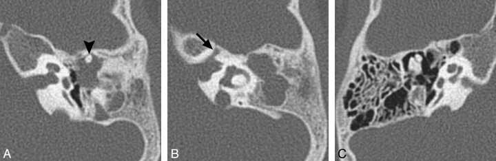
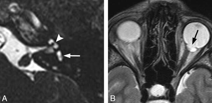
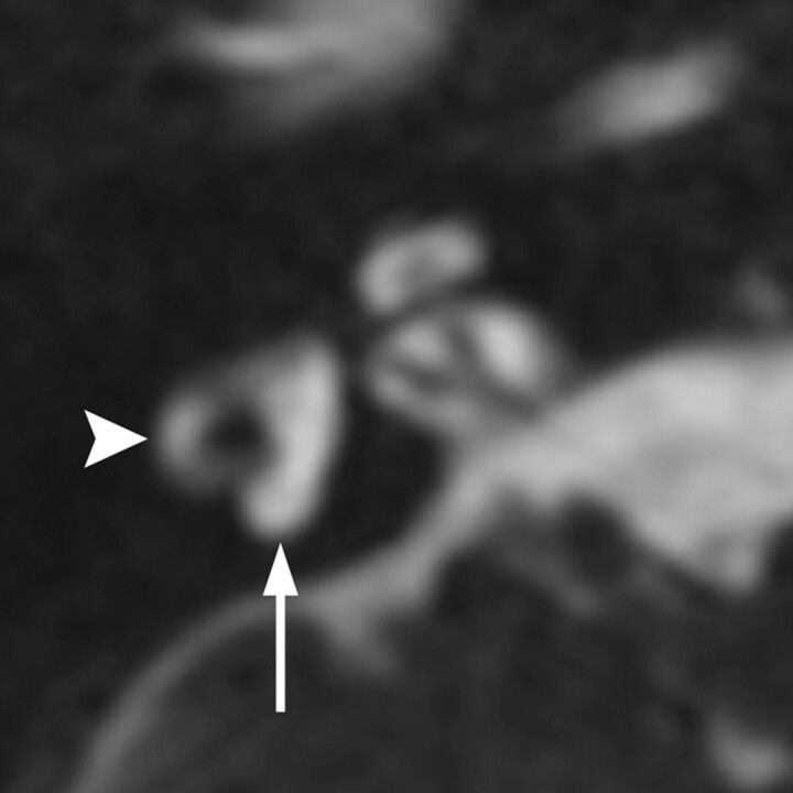
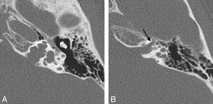
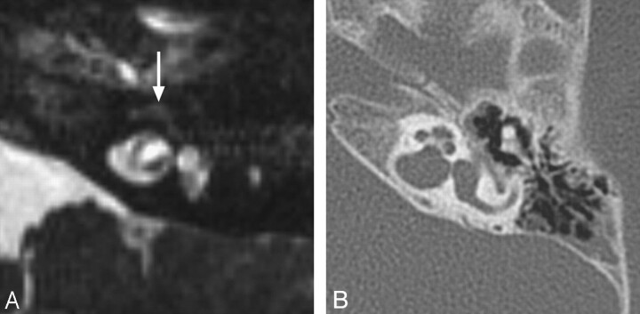
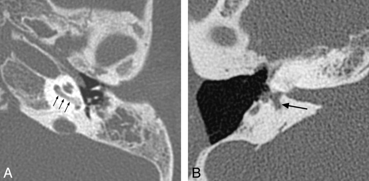
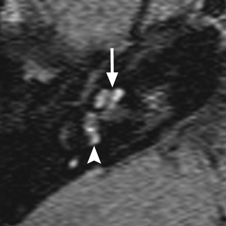
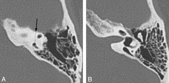
This article is the second in a 2-part series reviewing neuroimaging in childhood SNHL. Previously, we discussed the clinical work-up of children with hearing impairment, the classification of inner ear malformations, and congenital nonsyndromic causes of hearing loss. Here, we review and illustrate the most common syndromic hereditary and acquired causes of childhood SNHL, with an emphasis on entities that demonstrate inner ear abnormalities on cross-sectional imaging. Syndromes discussed include BOR syndrome, CHARGE syndrome, Pendred syndrome, Waardenburg syndrome, and X-linked hearing loss with stapes gusher. We conclude the article with a review of acquired causes of childhood SNHL, including infections, trauma, and neoplasms.


Figures

Fig 1.
Fig 1.
BOR syndrome. A, Axial CT image through the level of the cochlear aperture demonstrates a hypoplastic cochlea without a definable modiolus or apical turn. B, An image slightly superior to A demonstrates a medially displaced canal for the labyrinthine segment of the facial nerve (arrow) and an enlarged vestibular aqueduct. This patient also has a left middle ear cholesteatoma causing ossicular erosion. Only the head of the malleus remains (arrowhead in A). C, CT image through the right epitympanum in the same patient demonstrates a foreshortened dysplastic incus with an enlarged body that essentially articulates directly with the stapes.
Fig 2.
Fig 2.
CHARGE syndrome. A, Axial CISS image through the left temporal bone demonstrates a small vestibule (white arrow) and absence of the lateral SCC. The cochlea is also hypoplastic (arrowhead). B, Axial fast spin-echo T2WI through the brain and orbits demonstrates a left-sided coloboma (black arrow).
Fig 3.
Fig 3.
Waardenburg syndrome. Axial CISS image through the right temporal bone demonstrates absence of the posterior SCC (arrow) but a normal lateral SCC (arrowhead). This is a characteristic finding of Waardenburg and Alagille syndromes.
Fig 4.
Fig 4.
X-linked hearing loss with stapes gusher. A, Axial CT image through the mid-IAC on the left demonstrates a bulbous IAC, widening of the cochlear aperture, and cochlear hypoplasia with modiolar deficiency. The vestibule is also dilated. B, Axial CT image slightly superior to A demonstrates widening of the bony canal for the labyrinthine segment of the facial nerve (arrow).
Fig 5.
Fig 5.
Labyrinthine fibrosis. A, Axial CISS image through the left IAC demonstrates loss of normal high-fluid signal intensity in the left cochlea (arrow). B, On the corresponding axial CT image through the left temporal bone, the turns of the cochlea are not ossified; however, subtle thickening of the modiolus may reflect very early cochlear ossification.
Fig 6.
Fig 6.
Two cases of labyrinthitis ossificans. A, Axial left temporal bone image demonstrates increased attenuation in the basal turn of the cochlea (small arrows) in a patient with early labyrinthitis ossificans. B, Axial right temporal bone image in a patient who has undergone a previous tympanomastoidectomy demonstrates complete ossification of the cochlea and lateral and posterior SCCs, consistent with advanced labyrinthitis ossificans. Portions of the vestibule (large arrow) remain unossified. Note the normal-sized IAC, which helps to distinguish this from labyrinthine aplasia.
Fig 7.
Fig 7.
Autoimmune labyrinthitis. Axial gadolinium-enhanced T1WI through the right inner ear demonstrates enhancement of the cochlea (arrow), vestibule (arrowhead), and SCCs. Acute infectious labyrinthitis would have an identical appearance.
Fig 8.
Fig 8.
Transverse temporal bone fracture resulting in a perilymph fistula. A, Axial left temporal bone CT image through the level of the superior SCC demonstrates a transversely oriented fracture (arrow), which involves the posterior limb of the superior SCC. B, Axial CT image just inferior to A demonstrates pneumolabyrinth in the vestibule and cochlea. In addition, there is gas in the vestibular aqueduct, which happens to be enlarged in this patient.
Fig 9.
Fig 9.
Hemolabyrinth. Coronal unenhanced T1WI through the cochleae demonstrates increased signal intensity in the right cochlea (arrow), consistent with hemorrhage following trauma.
Fig 10.
Fig 10.
NF-2 axial gadolinium-enhanced T1WI through the level of the IACs demonstrates enhancing vestibular schwannomas in both IACs (arrows) and trigeminal schwannomas in both Meckel caves (arrowheads).
Fig 11.
Fig 11.
Pilocytic astrocytoma causing left-sided hearing loss. Axial T2WI demonstrates an exophytic hyperintense mass arising from the region of the left inferior cerebellar peduncle (arrow).

References

    1. Billings KR, Kenna MA. Causes of pediatric sensorineural hearing loss: yesterday and today. Arch Otolaryngol Head Neck Surg 1999; 125: 517– 21 - PubMed
    1. Lalwani AK, Castelein CM. Cracking the auditory genetic code: nonsyndromic hereditary hearing impairment. Am J Otol Jan 1999; 20: 115– 32 - PubMed
    1. Morton CC, Nance WE. Newborn hearing screening: a silent revolution. N Engl J Med 2006; 354: 2151– 64 - PubMed
    1. Kochhar A, Fischer SM, Kimberling WJ, et al. . Branchio-oto-renal syndrome. Am J Med Genet A 2007; 143A: 1671– 78 - PubMed
    1. Propst EJ, Blaser S, Gordon KA, et al. . Temporal bone findings on computed tomography imaging in branchio-oto-renal syndrome. Laryngoscope 2005; 115: 1855– 62 - PubMed