. 2011 May 20;332(6032):981-4.
doi: 10.1126/science.1202692.

Structures of the bacterial ribosome in classical and hybrid states of tRNA binding

Affiliations

Structures of the bacterial ribosome in classical and hybrid states of tRNA binding

Jack A Dunkle et al. Science. .

Abstract

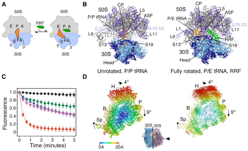
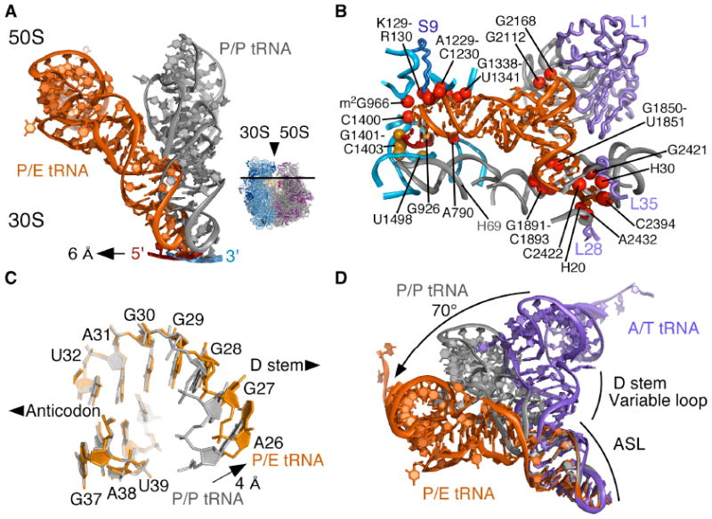
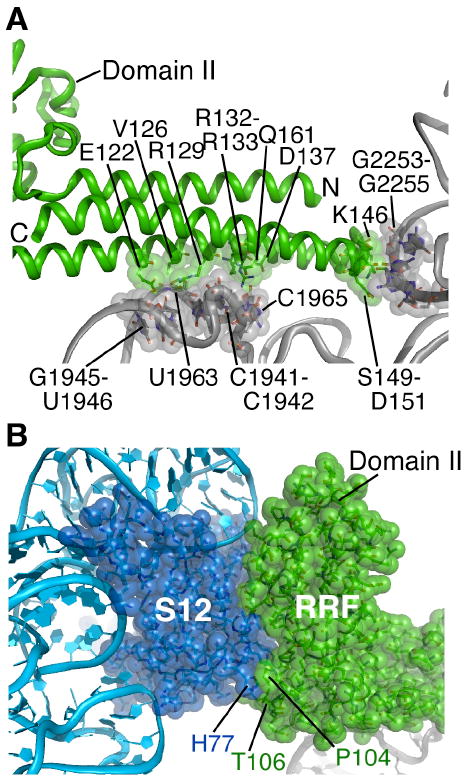
During protein synthesis, the ribosome controls the movement of tRNA and mRNA by means of large-scale structural rearrangements. We describe structures of the intact bacterial ribosome from Escherichia coli that reveal how the ribosome binds tRNA in two functionally distinct states, determined to a resolution of ~3.2 angstroms by means of x-ray crystallography. One state positions tRNA in the peptidyl-tRNA binding site. The second, a fully rotated state, is stabilized by ribosome recycling factor and binds tRNA in a highly bent conformation in a hybrid peptidyl/exit site. The structures help to explain how the ratchet-like motion of the two ribosomal subunits contributes to the mechanisms of translocation, termination, and ribosome recycling.


Figures

Fig. 1
Fig. 1
Ribosome recycling in bacteria and organelles. (A) Steps of ribosome recycling. After termination, ribosomes with deacylated tRNA in the P site undergo a structural rearrangement to a fully rotated state in which tRNA adopts a P/E hybrid state of binding and RRF is bound in the 50S P site. EF-G then catalyzes subunit dissociation (not shown). (B) Global views of the ribosome in a post-termination state (L) and intermediate state of recycling (R). The small subunit rRNA and proteins are colored light and dark blue, respectively, with the large subunit rRNA and proteins colored grey and magenta, respectively. Bound tRNA (orange), mRNA (dark red), and RRF (green) are also shown. (C) The dependence of subunit release on RRF, EF-G and GTP under crystallographic buffer conditions. Release was monitored by the loss of Cy5-labeled L1 fluorescence in 50S subunits from surface-immobilized ribosome complexes carrying Cy3-labeled tRNAPhe in the P site. Complexes imaged in the absence of factors (black diamonds) or in the presence of 10μM RRF (green circles), 20 μM EF-G/2mM GTP (pink inverted triangles), 10 μM RRF/20 μM EF-G/2mM GDPNP (blue triangles) or 10 μM RRF/20 μM EF-G/2mM GTP (red squares). Data reflect the mean +/- SD of normalized Cy5 fluorescence intensity as a function of time from three experimental replicates. (D) Conformational changes in the 70S ribosome during ratcheting. View of the 30S subunit from the perspective of the 50S subunit (inset). Shifts between equivalent RNA phosphorus atoms and protein Cα atoms in the unrotated (R0) and fully rotated (RF) states are color coded as indicated by the scale. Ribosomes were superimposed using the 50S subunit as the frame of reference (37). Difference vectors between equivalent phosphorus or Cα atoms of the 30S subunits in the unrotated and fully rotated ribosome structures are shown on the right.
Fig. 2
Fig. 2
Conformation of tRNA in the P/E hybrid state. (A) Movement of P/E tRNA and mRNA towards the E site when compared to P/P tRNA and mRNA. The direction of view is shown to the right. (B) View of mRNA and P/E tRNA interactions with the 30S subunit P site and 50S subunit E site. Residues that contact mRNA (gold) and P/E tRNA (red) are shown. Colors for the ribosome, mRNA and tRNA as in Fig. 1. (C) View of the P/E tRNA ASL/D stem junction (orange). P/P tRNA (grey) is shown for comparison, with an arrow indicating the widening of the helix major groove. (D) Comparison of ASL/D stem junctions between P/E tRNA (orange), P/P tRNA (grey), and A/T tRNA (purple). A/T tRNA structure is a homology model adapted from (12, 21). The bending angle for the A/T to P/E conformational change (70°) is shown.
Fig. 3
Fig. 3
Inter-subunit contacts in the fully rotated state. (A) Global view of inter-subunit contacts of the fully rotated state. Elements in each ribosomal subunit that contact rRNA in opposite subunit are color-coded red, while elements in each subunit that contact ribosomal proteins in the opposite subunit are color-coded gold. Ribosomal RNAs and proteins are colored as in Figure 1. Bridge numbering is adapted from (11, 25). The tip of helix H38 in bridge B1a is disordered in the present structures. (B) Bridge B3 serves as the pivot of inter-subunit rotation. The Mg2+ ion involved in inner-sphere coordination to the tandem sheared GA pairs in 16S rRNA and a fully hydrated Mg2+ ion in 23S rRNA are also shown. Ribosomal RNA colored as in Fig. 1. (C) Compression of helix H69 in 23S rRNA due to inter-subunit rotation. The direction of view is similar to Fig. 1. Color coding of the fully rotated ribosome (R) as in Fig. 1, with unrotated ribosome (U) in red. Nucleotide A1928 in 23S rRNA, nearly invariant in position, is shown for reference. (D) Movement of H68 due to disruption of A702 interactions and packing with P/E tRNA. Nucleotides involved in H68 packing with P/E tRNA are indicated. Elements of the fully rotated ribosome are colored as in Fig. 1. Elements of the unrotated ribosome are shown in red. Arrows indicate movement from the unrotated to fully rotated state. (E) Bridge B4 in the fully rotated state compared to that in state R0 (red). Residues involved in direct contact in the fully rotated state are shown. Coloring for the fully rotated state as in Fig. 1.
Fig. 4
Fig. 4
RRF interactions with the ribosome in the fully rotated state. (A) Contacts between RRF domain I and the P and A sites of the 50S subunit. Amino acids in RRF (green) and nucleotides in 23S rRNA (grey) in direct contact are shown. Helix H69 and the 30S subunit are behind the view shown. (B) Contacts between RRF and protein S12 in the 30S subunit. Amino acids at the junction of RRF domains I and II that interact closely with S12 are indicated. RRF, S12 and rRNAs colored as in Fig. 1.

References

    1. Schmeing TM, Ramakrishnan V. Nature. 2009;461:1234. - PubMed
    1. Munro JB, Sanbonmatsu KY, Spahn CM, Blanchard SC. Trends Biochem Sci. 2009;34:390. - PMC - PubMed
    1. Dunkle JA, Cate JH. Annu Rev Biophys. 2010;39:227. - PubMed
    1. Frank J, Agrawal RK. Nature. 2000;406:318. - PubMed
    1. Moazed D, Noller HF. Nature. 1989;342:142. - PubMed

Publication types

MeSH terms