. 2011 Jun 7;108(23):9589-94.
doi: 10.1073/pnas.1017945108. Epub 2011 May 19.

Canonical hedgehog signaling augments tumor angiogenesis by induction of VEGF-A in stromal perivascular cells

Affiliations

Canonical hedgehog signaling augments tumor angiogenesis by induction of VEGF-A in stromal perivascular cells

Weiwei Chen et al. Proc Natl Acad Sci U S A. .

Abstract

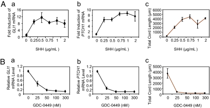
Hedgehog (Hh) signaling is critical to the patterning and development of a variety of organ systems, and both ligand-dependent and ligand-independent Hh pathway activation are known to promote tumorigenesis. Recent studies have shown that in tumors promoted by Hh ligands, activation occurs within the stromal microenvironment. Testing whether ligand-driven Hh signaling promotes tumor angiogenesis, we found that Hh antagonism reduced the vascular density of Hh-producing LS180 and SW480 xenografts. In addition, ectopic expression of sonic hedgehog in low-Hh-expressing DLD-1 xenografts increased tumor vascular density, augmented angiogenesis, and was associated with canonical Hh signaling within perivascular tumor stromal cells. To better understand the molecular mechanisms underlying Hh-mediated tumor angiogenesis, we established an Hh-sensitive angiogenesis coculture assay and found that fibroblast cell lines derived from a variety of human tissues were Hh responsive and promoted angiogenesis in vitro through a secreted paracrine signal(s). Affymetrix array analyses of cultured fibroblasts identified VEGF-A, hepatocyte growth factor, and PDGF-C as candidate secreted proangiogenic factors induced by Hh stimulation. Expression studies of xenografts and angiogenesis assays using combinations of Hh and VEGF-A inhibitors showed that it is primarily Hh-induced VEGF-A that promotes angiogenesis in vitro and augments tumor-derived VEGF to promote angiogenesis in vivo.


Conflict of interest statement

Conflict of interest statement: All authors are employees of Genentech, Inc., a wholly owned subsidiary of F. Hoffmann-La Roche Ltd., and may own equity in Roche.

Figures

Fig. 1.
Fig. 1.
Hh signaling promotes tumor angiogenesis in LS180 xenografts. (A) FACS analysis of cell-surface ligand expression in LS180 cells using the Hh antibody 5E1. (B) LS180 tumor growth during treatment with vehicle or the HPI GDC-0449 (100 mg/kg, orally, twice daily). Data are mean ± SD (n = 10/group). (C and D) Tumor vascular density analysis of LS180 xenografts treated with GDC-0449 (75 mg/kg, orally, twice daily) vs. vehicle following 3 d of drug administration. GDC-0449 reduces tumor relative vessel area (C; *P = 0.01) and relative vessel perimeter (D; *P = 0.03) compared with vehicle treatment (n = 10/group). The middle line of each diamond represents the mean, and the upper and lower borders of the diamonds the 95% confidence interval.
Fig. 2.
Fig. 2.
Canonical Hh signaling in stromal perivascular cells is associated with augmented tumor angiogenesis. (A) FACS analysis of cell-surface ligand expression in parental DLD-1 (Left) and in DLD-1.SHH vs. DLD-1.Vec cells (Right) using Hh antibody 5E1. (B) RT-PCR transcript comparisons for day 8 (D8) DLD-1.Vec vs. DLD-1.SHH tumors. Mouse Gli1 and Ptch1 transcripts were quantified using species-specific primers, and the mouse Rpl19 gene was used as an internal control for each animal. Data are mean ± SD (n = 10/group). (C Upper) Representative gross images of DLD-1.SHH tumors. Left-sided hash marks are separated by 1 mm. Note the enlarged, dark red appearance of the tumors. (Lower) H&E-stained section of DLD-1.SHH tumor showing numerous red blood-cell lakes (hemorrhages) marked by an asterisk (16). (D) Representative gross images (Upper) and H&E-stained section images (Lower) of DLD-1.Vec control tumors. (E–H) Isotopic in situ hybridization (ISH) analysis of mouse Ptch1 gene expression in D8 DLD-1.SHH tumor sections. (F and H) Insets of E and G showing Ptch1 up-regulation in stromal perivascular cells. Arrows highlight silver grains that identify sites of probe hybridization. Note that silver grains overlie cells surrounding tumor blood vessels (BV). (I and J) ISH analysis of Ptch1 expression in a DLD-1.Vec control tumor section. Few Ptch1 transcripts were detected adjacent to tumor blood vessels. (K and L) Vascular density analysis of D8 DLD-1.SHH (n = 9) and DLD-1.Vec (n = 10) xenografts. SHH overexpression in DLD-1 tumors significantly increased relative vessel area (*P = 0.006) and relative vessel perimeter (*P = 0.004). (Magnification: D, 70×; E, G, and I, 200×.)
Fig. 3.
Fig. 3.
Angiogenesis correlates with Hh target gene induction in myofibroblasts. (A) GLI1 and PTCH1 target gene induction in CCD-18Co myofibroblasts and angiogenesis are dosage sensitive to SHH. (A, a and b) RT-PCR analysis of GLI1 and PTCH1 in cultured CCD-18Co cells treated with increasing doses of SHH for 48 h. (A, c) Hh-mediated angiogenesis in CCD-18Co/HUVEC cocultures following increasing doses of SHH. Note that HUVEC cord formation, reflected by the total cord length, is dosage sensitive to SHH and mirrors Hh target gene induction in CCD-18Co cells. (B) GLI1 and PTCH1 target gene induction in CCD-18Co cells and angiogenesis are dosage sensitive to Hh inhibition. (B, a and b) RT-PCR analysis of GLI1 and PTCH1 in cultured CCD-18Co cells treated with 1 μg/mL SHH and increasing doses of GDC-0449 for 48 h. (B, c) Hh-mediated angiogenesis in CCD-18Co/HUVEC cocultures following increasing doses of GDC-0449. Note that HUVEC cord formation is dosage sensitive to GDC-0449 and mirrors the change in Hh target gene expression in CCD-18Co cells. Data are mean ± SD (n = 3).
Fig. 4.
Fig. 4.
VEGF-A is the predominant Hh-induced proangiogenic factor driving tumor angiogenesis in vitro and in vivo. (A) Conditioned media from SHH-stimulated CCD-18Co cells promotes HUVEC cord formation. Data are mean ± SD (n = 3). (B) U133P Affymetrix array analysis comparing gene expression of CCD-18Co cells cultured with vehicle vs. 1 μg/mL SHH for 72 h. GLI1 and PTCH1 were induced, as expected, as well as the genes of several secreted proangiogenic factors. Values represent the fold change in expression in SHH- vs. vehicle-treated CCD-18Co cells. The P values were obtained from all probes measuring a gene's transcripts. An asterisk signifies that similar fold changes were seen with RT-PCR analyses following 48 h of SHH exposure. (C and D) ISH analysis of Vegf-A expression in DLD-1.SHH tumors. Similar to mouse Ptch1 (Fig. 2 EH), Vegf-A is up-regulated in perivascular stromal cells in DLD-1.SHH tumors (D, arrows) Inset of C. BV locates blood vessels that are identified by their pink-red extracellular matrix and lumens. (E and F) ISH analysis in DLD-1.Vec control tumors shows little perivascular Vegf-A expression. (G) SHH-induced (1 μg/mL) in vitro angiogenesis was dosage sensitive to increasing concentrations of the α-VEGF-A antibody, G6-31. Data are mean ± SD (n = 3). (H) SHH-induced angiogenesis was abolished by 66.7 nM G6-31, indicating that SHH-induced angiogenesis in this model system is VEGF dependent. Data are mean ± SD (n = 3). (I) Vascular density analysis of Hh-producing SW480 xenograft tumors following 6 d of treatment with either HPI Hh-Antag (100 mg/kg, orally, twice daily), G6-31 (5 mg/kg, i.p., 2×/week), or a combination of both (n = 10/group). *P = 0.026, **P = 0.0001, ***P = 0.0002. No significant difference was seen between the G6-31 and combination group (P = 0.38). (Magnification: C and E, 280×.)

References

    1. Scales SJ, de Sauvage FJ. Mechanisms of Hedgehog pathway activation in cancer and implications for therapy. Trends Pharmacol Sci. 2009;30:303–312. - PubMed
    1. Yauch RL, et al. A paracrine requirement for hedgehog signalling in cancer. Nature. 2008;455:406–410. - PubMed
    1. Tian H, et al. Hedgehog signaling is restricted to the stromal compartment during pancreatic carcinogenesis. Proc Natl Acad Sci USA. 2009;106:4254–4259. - PMC - PubMed
    1. Byrd N, Grabel L. Hedgehog signaling in murine vasculogenesis and angiogenesis. Trends Cardiovasc Med. 2004;14:308–313. - PubMed
    1. Olive KP, et al. Inhibition of Hedgehog signaling enhances delivery of chemotherapy in a mouse model of pancreatic cancer. Science. 2009;324:1457–1461. - PMC - PubMed

MeSH terms

Associated data