. 2011 Sep;25(9):2921-36.
doi: 10.1096/fj.11-183798. Epub 2011 May 19.

Intrinsic apoptosis in mechanically ventilated human diaphragm: linkage to a novel Fos/FoxO1/Stat3-Bim axis

Affiliations

Intrinsic apoptosis in mechanically ventilated human diaphragm: linkage to a novel Fos/FoxO1/Stat3-Bim axis

Huibin Tang et al. FASEB J. 2011 Sep.

Abstract

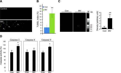
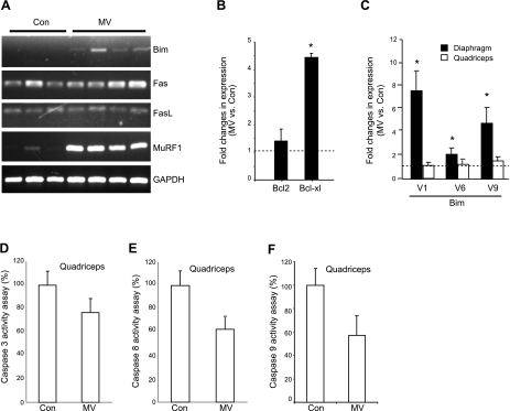
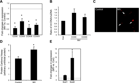
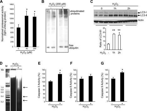
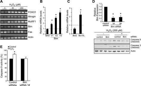
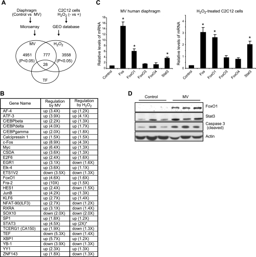
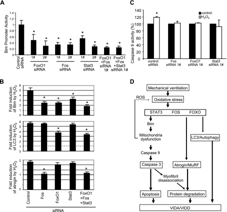
Mechanical ventilation (MV) is a life-saving measure in many critically ill patients. However, prolonged MV results in diaphragm dysfunction that contributes to the frequent difficulty in weaning patients from the ventilator. The molecular mechanisms underlying ventilator-induced diaphragm dysfunction (VIDD) remain poorly understood. We report here that MV induces myonuclear DNA fragmentation (3-fold increase; P<0.01) and selective activation of caspase 9 (P<0.05) and Bcl2-interacting mediator of cell death (Bim; 2- to 7-fold increase; P<0.05) in human diaphragm. MV also statistically significantly down-regulates mitochondrial gene expression and induces oxidative stress. In cultured muscle cells, we show that oxidative stress activates each of the catabolic pathways thought to underlie VIDD: apoptotic (P<0.05), proteasomal (P<0.05), and autophagic (P<0.01). Further, silencing Bim expression blocks (P<0.05) oxidative stress-induced apoptosis. Overlapping the gene expression profiles of MV human diaphragm and H₂O₂-treated muscle cells, we identify Fos, FoxO1, and Stat3 as regulators of Bim expression as well as of expression of the catabolic markers atrogin and LC3. We thus identify a novel Fos/FoxO1/Stat3-Bim intrinsic apoptotic pathway and establish the centrality of oxidative stress in the development of VIDD. This information may help in the design of specific drugs to prevent this condition.


Figures

Figure 1.
Figure 1.
MV results in DNA fragmentation and activates caspase 3 and 9 in human diaphragm. A) TUNEL staining was performed on cryosections of control and ventilated human diaphragm. Fragmented genomic DNA was labeled with FITC-conjugated dUTP; positive signals appear green. Myonuclei are visualized by DAPI staining (blue). Note that the positive TUNEL staining signals (green) are localized in nuclei stained by DAPI (blue). B) TUNEL-positive nuclei were counted and normalized to total myonuclei; ratio is shown as the TUNEL index. Control (Con), n = 9; MV, n = 10. C) DNA fragmentation was measured by PCR-based detection and visualized by electrophoresis in 2% agarose gel. Density of the PCR products was quantitated with ImageJ and normalized to the total DNA input. M, DNA markers. Control, n = 5; MV, n = 8. D) Caspase 8 and 9 enzymatic activities from control and MV human diaphragm lysate were measured by fluorometric assay. Results are presented as relative fluorescence units after normalization to total protein amount. Control, n = 7; MV, n = 9. *P < 0.05; **P < 0.01.
Figure 2.
Figure 2.
MV transcriptionally up-regulates molecular markers of intrinsic apoptosis in human diaphragm but not in quadriceps muscle. A) Total RNA was extracted from human diaphragm, and RT-PCR was performed and visualized on agarose gel to show the expression changes in apoptotic markers and the ubiquitin ligase MuRF1. B) Quantitative real-time PCR was performed on the ventilated and control human diaphragm. Transcriptional levels of the Bcl2 family members were examined. Control (Con), n = 7; MV, n = 9. C) Quantitative real-time PCR was performed on the ventilated and control human diaphragm, as well as the nonrespiratory quadriceps muscle. Transcriptional expression levels of the Bim splicing variants were examined. Note that changes in expression of the Bim splice variants with ventilation occur in diaphragm but not in nonrespiratory quadriceps muscle. Control, n = 8; MV, n = 7. D–F) Enzymatic activities of caspases were measured in nonrespiratory quadriceps muscles from ventilated and control subjects. Note that MV did not induce the activities of caspase 3 (D), caspase 8 (E), or caspase 9 (F) in quadriceps muscle. Control, n = 8; MV, n = 7. *P < 0.05.
Figure 3.
Figure 3.
MV induces oxidative stress and impairs mitochondrial function in human diaphragm. A) Total RNA was extracted from ventilated and control human diaphragm, and quantitative real-time PCR was performed. Fold changes of COX gene expression levels were calculated as MV over control after normalization to γ-actin levels. Note that expression levels of COX I, II, III, and IV genes in ventilated human diaphragm are reduced. Control (Con), n = 7; MV, n = 9. B) DNA from ventilated and control human diaphragm was extracted, and real-time PCR was performed to examine the levels of the genomic DNA of the β-actin gene as well as the mitochondrial DNA levels of the COX I and II genes. Ratio of the COX I or II DNA levels vs. β-actin nuclear DNA levels was used to indicate relative mitochondrial content. Note that mitochondrial DNA content does not change with MV. Control, n = 5; MV, n = 5. P = 0.15. C) DHE staining was performed on cryosections of ventilated and control human diaphragm. Positive DHE staining shows as red (arrows). D) Carbonyl assay was performed on protein extracts from ventilated and control human diaphragm. Light unit of the carbonyl assay result was normalized to total protein input. Control, n = 7; MV, n = 9. E) Real-time PCR was used to quantitate the expression levels of the cytosolic and mitochondrial antioxidant genes, SOD1 and SOD2, respectively. Note that only SOD2 is significantly elevated with MV. Control, n = 7; MV, n = 9. *P < 0.05.
Figure 4.
Figure 4.
Oxidative stress is sufficient to activate the UPS, autophagic, and intrinsic apoptotic pathways. A) Differentiated C2C12 myotubes were treated with H2O2 for 24 h, and proteasomal activity was measured in the cell lysate. Measured light unit was normalized to the total protein level. n = 6. B) C2C12 myotubes were treated with H2O2 for 24 h, and equal amounts of total protein extracts were run with PAGE (4–12%). Polyubiquitinated proteins were visualized by Western blot analysis with anti-ubiquitin antibody. Note that the free ubiquitin level did not change significantly. C) C2C12 cells were treated with H2O2 (1 mM), and cell lysates were subjected to Western blot analysis with anti-LC3 antibody. Total input protein levels were 20 μg, and the α-actin protein level was also detected for normalization. Protein expression levels of LC3-I and II were quantitated by ImageJ and normalized to the actin protein level. Ratio of LC3-II to LC3-I is significantly induced. n = 3. D) Genomic DNA was extracted from H2O2 (1 mM, 24 h)-treated C2C12 myotubes and visualized with 2% agarose gel. Representative gel is shown. M, DNA marker. Arrows indicate DNA fragments. E–G) Caspase 3 (D), 8 (E), and 9 (F) activities were assayed on C2C12 cell lysates, with and without H2O2 treatment (200 μM, 24 h). n = 6. *P < 0.05; **P < 0.01.
Figure 5.
Figure 5.
Oxidative stress-induced Bim is required for the activation of intrinsic apoptosis in cultured muscle cells. A) Differentiated C2C12 myotubes were treated with H2O2 for 24 h. Total RNA was extracted from myotubes for RT-PCR reaction, and PCR products were visualized by running with 1.5% agarose gel and stained by ethidium bromide. B, C) Quantitative real-time PCR was performed on H2O2-treated C2C12 muscle cells. γ-Actin was used for normalization. Each sample was run in triplicate; fold change was calculated by normalizing the H2O2-treated to the nontreated myotube samples. n = 3. D) Control and Bim siRNAs were transfected into C2C12 muscle cells, which were then induced to differentiate into myotubes and treated with H2O2 (200 μM) for 24 h. Real-time PCR was used to quantitate the efficiency of siRNAs (top), and Western blot analysis was performed to show the level of cleaved/activated caspase 3 and 9 (bottom) in the presence or absence of Bim siRNA. n = 3. E) C2C12 cells was transfected with control and Bim siRNAs for 3 d, and 200 μM H2O2 was added for 24 h. Cell lysates were collected for caspase 9 activity assay by a Caspase-Glo assay system and normalized to the total protein input. n = 6. *P < 0.05.
Figure 6.
Figure 6.
Systemic search for transcription factors regulated in common by oxidative stress within the in vitro and in vivo models. A) Microarray analysis was performed on RNA samples from control and human MV diaphragm, and data were analyzed by GeneSpring GX11. Raw data on H2O2-treated C2C12 cells from the GEO database in the NCBI GenBank were analyzed by GeneSpring GX11. With cutoff set at P < 0.05, commonly regulated entities were mined as shown in Venn diagram. GeneGo software was then used to identify known transcription factors for these commonly regulated genes. Note that 28 nonredundant transcription factors were identified. B) Transcription factors regulated in common in both MV human diaphragm and H2O2-treated muscle cells. *Gene was not found on microarray but was confirmed by real-time PCR. C) Real-time PCR was performed on RNA from MV human diaphragm and H2O2-treated muscle cells; fold changes were calculated by normalizing to their respective controls (diaphragm control, n=7; MV, n=9; C2C12 cells, n=3). *P < 0.05. D) Total proteins from human diaphragm (control and ventilated, n=3) were extracted; Western blot analysis was performed. Actin protein was detected as the loading control.
Figure 7.
Figure 7.
Fos, FoxO1, and Stat3-mediated regulation of autophagy, atrophy, and apoptosis in response to oxidative stress. A) C2C12 cells were transfected with siRNAs, together with a Bim promoter reporter construct and β-Gal reporter construct. Reporter gene assay was performed 3 d later; luciferase light unit was normalized to the cotransfected β-Gal activity. Note that two different siRNAs (1# and 2#) against different regions of each gene were used to minimize the potential off-target effect. n = 3. B) Regulatory effect of Fos, FoxO1, and Stat3 on atrophic, autophagic, and apoptotic markers in response to oxidative stress. C2C12 cells were transfected with siRNAs for 3 d and treated with H2O2 (200 μM) for 24 h on d 4. Total RNA was collected, and real-time PCR was performed to examine the gene expression levels. Note that all three transcription factors inhibit Bim gene expression in response to oxidative stress, but Fos also regulates atrogin, and FoxO1 regulates LC3. n = 4. C) Fos, FoxO1, and Stat3 are required for H2O2-dependent activation of apoptosis. siRNAs against Fos, FoxO1, and Stat3 were transfected into C2C12 cells for 3 d and treated with H2O2 (200 mM) for 24 h. Caspase 9 activity was measured and normalized to the total protein input. n = 3. D) Schematic diagram of the proposed molecular mechanisms underlying MV-induced diaphragm atrophy and dysfunction. See detailed description in text. *P < 0.05.

Similar articles

Cited by

References

    1. Gayan-Ramirez G., Decramer M. (2002) Effects of mechanical ventilation on diaphragm function and biology. Eur. Respir. J. 20, 1579–1586 - PubMed
    1. Laghi F., Cattapan S., Jubran A., Parthasarathy S., Warshawsky P., Choi Y., Tobin M. (2003) Is weaning failure caused by low-frequency fatigue of the diaphragm? Am. J. Respir. Crit. Care Med. 167, 120–127 - PubMed
    1. Swartz M., Marino P. (1985) Diaphragmatic strength during weaning from mechanical ventilation. Chest 88, 736–739 - PubMed
    1. Alía I., Esteban A. (2000) Weaning from mechanical ventilation. Crit. Care 4, 72–80 - PMC - PubMed
    1. Esteban A., Frutos F., Tobin M., Alía I., Solsona J., Valverdú I., Fernández R., de la Cal M., Benito S., Tomás R. (1995) A comparison of four methods of weaning patients from mechanical ventilation. Spanish Lung Failure Collaborative Group. N. Engl. J. Med. 332, 345–350 - PubMed

Publication types

MeSH terms