

Epigenetic regulation of early osteogenesis and mineralized tissue formation by a HOXA10-PBX1-associated complex
- PMID: 21597276
- PMCID: PMC3178072
- DOI: 10.1159/000324790
Epigenetic regulation of early osteogenesis and mineralized tissue formation by a HOXA10-PBX1-associated complex
Abstract
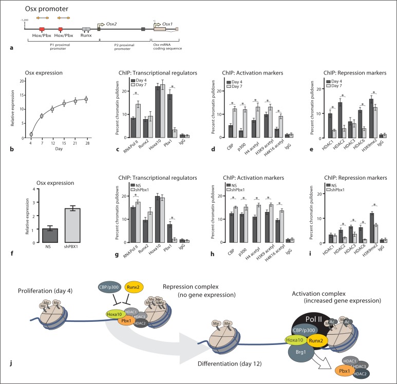
Homeodomain-containing (HOX) factors such as the abdominal class homeodomain protein HOXA10 and the TALE-family protein PBX1 form coregulatory complexes and are potent transcriptional and epigenetic regulators of tissue morphogenesis. We have identified that HOXA10 and PBX1 are expressed in osteoprogenitors; however, their role in osteogenesis has not been established. To determine the mechanism of HOXA10-PBX-mediated regulation of osteoblast commitment and the related gene expression, PBX1 or HOX10 were depleted (shRNA or genetic deletion, respectively) or exogenously expressed in C3H10T1/2, bone marrow stromal progenitors, and MC3T3-E1 (preosteoblast) cells. Overexpression of HOXA10 increased the expression of osteoblast-related genes, osteoblast differentiation and mineralization; expression of PBX1 impaired osteogenic commitment of pluripotent cells and the differentiation of osteoblasts. In contrast, the targeted depletion of PBX1 by shRNA increased the expression of bone marker genes (osterix, alkaline phosphatase, BSP, and osteocalcin). Chromatin-associated PBX1 and HOXA10 were present at osteoblast-related gene promoters preceding gene expression, but PBX1 was absent from promoters during the transcription of bone-related genes, including osterix (Osx). Further, PBX1 complexes were associated with histone deacetylases normally linked with chromatin inactivation. Loss of PBX1 but not of HOXA10 from the Osx promoter was associated with increases in the recruitment of histone acetylases (p300), as well as decreased H3K9 methylation, reflecting transcriptional activation. We propose PBX1 plays a central role in attenuating the activity of HOXA10 as an activator of osteoblast-related genes and functions to establish the proper timing of gene expression during osteogenesis, resulting in proper matrix maturation and mineral deposition in differentiated osteoblasts.
Copyright © 2011 S. Karger AG, Basel.
Figures

Similar articles
-
Pbx1 represses osteoblastogenesis by blocking Hoxa10-mediated recruitment of chromatin remodeling factors.Mol Cell Biol. 2010 Jul;30(14):3531-41. doi: 10.1128/MCB.00889-09. Epub 2010 May 3. Mol Cell Biol. 2010. PMID: 20439491 Free PMC article.
-
HOXA10 controls osteoblastogenesis by directly activating bone regulatory and phenotypic genes.Mol Cell Biol. 2007 May;27(9):3337-52. doi: 10.1128/MCB.01544-06. Epub 2007 Feb 26. Mol Cell Biol. 2007. PMID: 17325044 Free PMC article.
-
Transcriptional regulation of bone sialoprotein gene expression by Osx.Biochem Biophys Res Commun. 2016 Aug 5;476(4):574-579. doi: 10.1016/j.bbrc.2016.05.164. Epub 2016 May 31. Biochem Biophys Res Commun. 2016. PMID: 27261434
-
The developmental control of osteoblast-specific gene expression: role of specific transcription factors and the extracellular matrix environment.Crit Rev Oral Biol Med. 1999;10(1):40-57. doi: 10.1177/10454411990100010201. Crit Rev Oral Biol Med. 1999. PMID: 10759426 Review.
-
The TALE never ends: A comprehensive overview of the role of PBX1, a TALE transcription factor, in human developmental defects.Hum Mutat. 2022 Sep;43(9):1125-1148. doi: 10.1002/humu.24388. Epub 2022 May 18. Hum Mutat. 2022. PMID: 35451537 Review.
Cited by
-
Role of Hox genes in stem cell differentiation.World J Stem Cells. 2015 Apr 26;7(3):583-95. doi: 10.4252/wjsc.v7.i3.583. World J Stem Cells. 2015. PMID: 25914765 Free PMC article. Review.
-
The RUNX2 cistrome in osteoblasts: characterization, down-regulation following differentiation, and relationship to gene expression.J Biol Chem. 2014 Jun 6;289(23):16016-31. doi: 10.1074/jbc.M114.552216. Epub 2014 Apr 24. J Biol Chem. 2014. PMID: 24764292 Free PMC article.
-
Mediation by Placental DNA Methylation of the Association of Prenatal Maternal Smoking and Birth Weight.Am J Epidemiol. 2019 Nov 1;188(11):1878-1886. doi: 10.1093/aje/kwz184. Am J Epidemiol. 2019. PMID: 31497855 Free PMC article.
-
A hydrophobic residue in the TALE homeodomain of PBX1 promotes epithelial-to-mesenchymal transition of gastric carcinoma.Oncotarget. 2017 Jul 18;8(29):46818-46833. doi: 10.18632/oncotarget.17473. Oncotarget. 2017. PMID: 28514754 Free PMC article.
-
Baf45a Mediated Chromatin Remodeling Promotes Transcriptional Activation for Osteogenesis and Odontogenesis.Front Endocrinol (Lausanne). 2022 Jan 3;12:763392. doi: 10.3389/fendo.2021.763392. eCollection 2021. Front Endocrinol (Lausanne). 2022. PMID: 35046892 Free PMC article.
References
-
- Lu X., Gilbert L., He X., Rubin J., Nanes M.S. Transcriptional regulation of the osterix (Osx, Sp7) promoter by tumor necrosis factor identifies disparate effects of mitogen-activated protein kinase and NF kappa B pathways. J Biol Chem. 2006;281:6297–6306. - PubMed
Publication types
MeSH terms
Substances
Grants and funding
LinkOut - more resources
Full Text Sources
Research Materials
Miscellaneous
