. 2011 Jul 1;10(13):2162-71.
doi: 10.4161/cc.10.13.16238. Epub 2011 Jul 1.

Cellular studies reveal mechanistic differences between taccalonolide A and paclitaxel

Affiliations

Cellular studies reveal mechanistic differences between taccalonolide A and paclitaxel

April L Risinger et al. Cell Cycle. .

Abstract

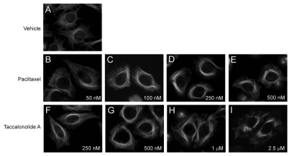
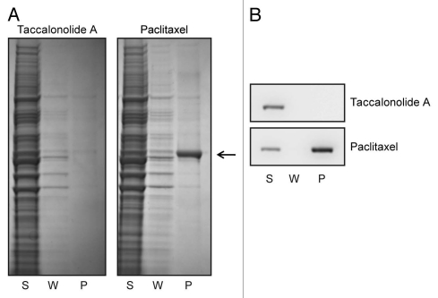
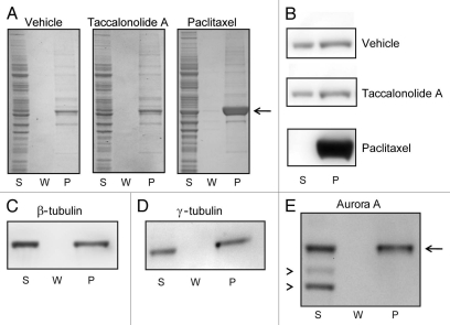
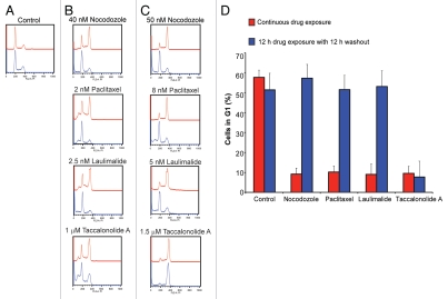
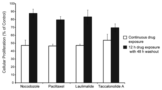
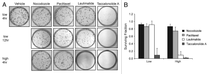
Taccalonolide A is a microtubule stabilizer that has cellular effects almost identical to paclitaxel. However, biochemical studies show that, unlike paclitaxel, taccalonolide A does not enhance purified tubulin polymerization or bind tubulin/microtubules. Mechanistic studies aimed at understanding the nature of the differences between taccalonolide A and paclitaxel were conducted. Our results show that taccalonolide A causes bundling of interphase microtubules at concentrations that cause antiproliferative effects. In contrast, the concentration of paclitaxel that initiates microtubule bundling is 31-fold higher than its IC 50. Taccalonolide A's effects are further differentiated from paclitaxel in that it is unable to enhance the polymerization of tubulin in cellular extracts. This finding extends previous biochemical results with purified brain tubulin to demonstrate that taccalonolide A requires more than tubulin and a full complement of cytosolic proteins to cause microtubule stabilization. Reversibility studies were conducted and show that the cellular effects of taccalonolide A persist after drug washout. In contrast, other microtubule stabilizers, including paclitaxel and laulimalide, demonstrate a much higher degree of cellular reversibility in both short-term proliferation and long-term clonogenic assays. The propensity of taccalonolide A to alter interphase microtubules at antiproliferative concentrations as well as its high degree of cellular persistence may explain why taccalonolide A is more potent in vivo than would be expected from cellular studies. The close linkage between the microtubule bundling and antiproliferative effects of taccalonolide A is of interest given the recent hypothesis that the effects of microtubule targeting agents on interphase microtubules might play a prominent role in their clinical anticancer efficacy.


Figures

Figure 1
Figure 1
Effects of paclitaxel or taccalonolide A on interphase microtubules in HeLa cells. Cells were incubated with vehicle (top row) or the indicated concentrations of paclitaxel (middle row) or taccalonolide A (bottom row) for 18 h. Microtubules were visualized by indirect immunofluorescence of β-tubulin.
Figure 2
Figure 2
Taccalonolide A is unable to form cold stable microtubules in cell lysates. HeLa cell lysates were collected, chilled to depolymerize microtubules and then treated with 20 µM paclitaxel or 20 µM taccalonolide A for 5 min at 37°C to reform microtubules. Lysates were then re-chilled to evaluate the ability of the stabilizers to initiate the formation of cold-stable microtubules. Microtubule polymer was pelleted by centrifugation and soluble tubulin heterodimers remained in the supernatant. The total protein (A) and β-tubulin (B) levels present in the supernatant (S), wash (W) and pellet (P) fractions were evaluated by total protein staining or β-tubulin immunoblotting, respectively. The location of tubulin in the total protein stained gel is indicated with an arrow.
Figure 3
Figure 3
Effects of paclitaxel or taccalonolide A on tubulin polymer formation in cytosolic extracts. HeLa cell lysates were collected, chilled to depolymerize pre-existing microtubules and then incubated with vehicle, 20 µM paclitaxel or 20 µM taccalonolide A for 30 min at 37°C. Microtubule polymer was separated from soluble tubulin by centrifugation at room temperature. The total protein (A) and β-tubulin (B) levels present in the supernatant (S), wash (W) and pellet (P) were determined by total protein staining or β-tubulin immunoblotting, respectively. The location of tubulin in the total protein stained gel is indicated with an arrow (3A). β-tubulin (C) and the microtubule associated proteins γ-tubulin (D) and Aurora A (E, arrow), were detected in the microtubule containing pellet (P) of samples treated with 100 µM taccalonolide A as compared to non-specific background bands, which were retained in the supernatant (E, arrowheads).
Figure 4
Figure 4
Reversibility of the cell cycle block induced by microtubule disrupting agents. Cell cycle profile of HeLa cells 12 h after drug addition (upper, red tracing in each part) or after an additional 12 h of drug washout (lower, blue tracing in each part). (A) Normal cell cycle distribution of HeLa cells. (B) Concentrations of each drug that caused significant but incomplete G2/M arrest. (C) Concentrations of each drug that caused G2/M accumulation of the majority of cells. (D) Quantitation of the percentage of cells from (C) that were in G1 after 12 h drug exposure or subsequent washout depicted in red and blue, respectively.
Figure 5
Figure 5
Antiproliferative effects of continuous or 12 h drug exposure. The concentration of drug required to observe a 50% inhibition in cellular proliferation following continuous 60 h incubation was determined (white bars). The antiproliferative effects of these same concentrations were evaluated after a 12 h exposure followed by drug washout for an additional 48 h (black bars).
Figure 6
Figure 6
Effects of 4 and 12 h drug exposure on clonogenic cell viability. (A) HeLa cells were treated with vehicle or concentrations of drug that inhibited cellular proliferation by 50% (low) or that caused G2/M arrest (high) for 4 or 12 h before media was replaced. Colony formation was determined after an additional 10 days of cell growth. (B) the surviving fraction of colony forming cells after a 4 h drug treatment and subsequent washout as compared to vehicle treated controls.

Comment in

References

    1. Dumontet C, Jordan MA. Microtubule-binding agents: a dynamic field of cancer therapeutics. Nat Rev Drug Discov. 2010;9:790–803. - PMC - PubMed
    1. Fojo T, Menefee M. Mechanisms of multidrug resistance: the potential role of microtubule-stabilizing agents. Ann Oncol. 2007;18(Suppl. 5):v3–v8. - PubMed
    1. Pusztai L. Markers predicting clinical benefit in breast cancer from microtubule-targeting agents. Ann Oncol. 2007;18(Suppl. 12):xii15–xii20. - PubMed
    1. Galsky MD, Dritselis A, Kirkpatrick P, Oh WK. Cabazitaxel. Nat Rev Drug Discov. 2010;9:677–678. - PubMed
    1. Morris PG, Fornier MN. Microtubule active agents: beyond the taxane frontier. Clin Cancer Res. 2008;14:7167–7172. - PubMed

Publication types

MeSH terms

LinkOut - more resources