Review
. 2011 Sep;18(9):1478-86.
doi: 10.1038/cdd.2011.65. Epub 2011 May 20.

Redox modulation by S-nitrosylation contributes to protein misfolding, mitochondrial dynamics, and neuronal synaptic damage in neurodegenerative diseases

Affiliations
Review

Redox modulation by S-nitrosylation contributes to protein misfolding, mitochondrial dynamics, and neuronal synaptic damage in neurodegenerative diseases

T Nakamura et al. Cell Death Differ. 2011 Sep.

Abstract

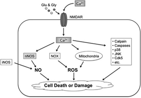
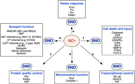
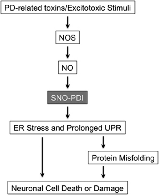
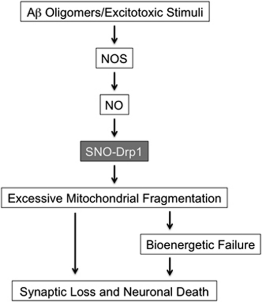
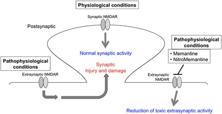
The pathological processes of neurodegenerative disorders such as Alzheimer's and Parkinson's diseases engender synaptic and neuronal cell damage. While mild oxidative and nitrosative (nitric oxide (NO)-related) stress mediates normal neuronal signaling, excessive accumulation of these free radicals is linked to neuronal cell injury or death. In neurons, N-methyl-D-aspartate (NMDA) receptor (NMDAR) activation and subsequent Ca(2+) influx can induce the generation of NO via neuronal NO synthase. Emerging evidence has demonstrated that S-nitrosylation, representing covalent reaction of an NO group with a critical protein thiol, mediates the vast majority of NO signaling. Analogous to phosphorylation and other posttranslational modifications, S-nitrosylation can regulate the biological activity of many proteins. Here, we discuss recent studies that implicate neuropathogenic roles of S-nitrosylation in protein misfolding, mitochondrial dysfunction, synaptic injury, and eventual neuronal loss. Among a growing number of S-nitrosylated proteins that contribute to disease pathogenesis, in this review we focus on S-nitrosylated protein-disulfide isomerase (forming SNO-PDI) and dynamin-related protein 1 (forming SNO-Drp1). Furthermore, we describe drugs, such as memantine and newer derivatives of this compound that can prevent both hyperactivation of extrasynaptic NMDARs as well as downstream pathways that lead to nitrosative stress, synaptic damage, and neuronal loss.


Figures

Figure 1
Figure 1
Excessive stimulation of NMDARs triggers intracellular signaling pathways leading to synaptic damage and neuronal cell loss. Hyperactivation of NMDARs by glutamate (Glu) and glycine (Gly) induces excessive Ca2+ influx and activation of nNOS leading to NO production. Ca2+ signaling also promotes ROS generation via activation of NADPH oxidase and dysfunctional mitochondria. iNOS, which is predominantly present in astrocytes, can produce toxic levels of NO, representing NMDAR-independent NO synthesis. In addition to producing RNS and ROS, NMDAR/Ca2+ signaling influences the activity of many other proteins (examples are listed at right), contributing to synaptic dysfunction and neuronal death
Figure 2
Figure 2
Representative examples of S-nitrosylated proteins that can regulate neuronal function. NO generated in the nervous system modulates the activity of various proteins via S-nitrosylation (or further oxidation), controlling (a) redox responses, (b) synaptic function, (c) protein quality control, (d) mitochondrial function, (e) transcriptional regulation, and (f) cell death and injury. Depending on the levels of NO, S-nitrosylation of these proteins can mediate either neuroprotective or neurodestructive pathways
Figure 3
Figure 3
Possible signaling pathways whereby SNO-PDI contributes to protein misfolding and neuronal damage. In PD, mitochondrial dysfunction caused by pesticides, herbicides, or other environmental toxins can trigger NO and ROS production, possibly via mitochondrial pathways and the NMDAR/Ca2+ cascade., , , NO produced by NOS reacts with sulfhydryl groups of PDI to form SNO-PDI, inhibiting its isomerase and chaperone activities. SNO-PDI formation causes ER stress and a prolonged UPR, and thereby contributes to neuronal cell injury, in part, by triggering accumulation of misfolded proteins
Figure 4
Figure 4
Possible signaling pathways that S-nitrosylated Drp1 contribute to excessive mitochondrial fission and synaptic damage. Soluble oligomers of Aβ peptide, thought to be a key mediator in AD pathogenesis, can facilitate neuronal NO and ROS production in both NMDAR-dependent and -independent manners., , S-Nitrosylation of Drp1 (forming SNO-Drp1) causes excessive mitochondrial fragmentation in neurodegenerative conditions. Compromised mitochondrial dynamics may result in the impairment of bioenergetics, and thus contribute to synaptic damage and eventually neuronal cell death
Figure 5
Figure 5
‘UFO'-type neuroprotective drugs, like memantine and NitroMemantine, preferentially block extrasynaptic NMDARs. Physiological synaptic activity of the NMDAR is required for neurotransmission and neuronal cell survival. In contrast, excessive activation of extrasynaptic NMDARs induces synaptic injury and neuronal loss, and is often associated with the accumulation of misfolded proteins as well as mitochondrial dysfunction. Conventional NMDAR antagonists, such as MK-801, completely block all receptor activity, including physiological synaptic activity, and thus result in severe side effects and clinical intolerability. Memantine and the newer NitroMemantine drugs preferentially block excessive (pathological) extrasynaptic NMDA receptor activity, while relatively sparing normal (physiological) synaptic activity,

References

    1. Beal MF. Experimental models of Parkinson's disease. Nat Rev Neurosci. 2001;2:325–334. - PubMed
    1. Lipton SA, Rosenberg PA. Excitatory amino acids as a final common pathway for neurologic disorders. N Engl J Med. 1994;330:613–622. - PubMed
    1. Lipton SA. Paradigm shift in neuroprotection by NMDA receptor blockade: memantine and beyond. Nat Rev Drug Discov. 2006;5:160–170. - PubMed
    1. Garthwaite J, Charles SL, Chess-Williams R. Endothelium-derived relaxing factor release on activation of NMDA receptors suggests role as intercellular messenger in the brain. Nature. 1988;336:385–388. - PubMed
    1. Brennan AM, Suh SW, Won SJ, Narasimhan P, Kauppinen TM, Lee H, et al. NADPH oxidase is the primary source of superoxide induced by NMDA receptor activation. Nat Neurosci. 2009;12:857–863. - PMC - PubMed

Publication types

Substances