. 2011 Aug;32(1):31-8.
doi: 10.1007/s10974-011-9247-8. Epub 2011 May 20.

Generation of a monoclonal antibody reactive to prefusion myocytes

Affiliations

Generation of a monoclonal antibody reactive to prefusion myocytes

Tomohiro Kurisaki et al. J Muscle Res Cell Motil. 2011 Aug.

Abstract

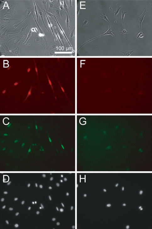
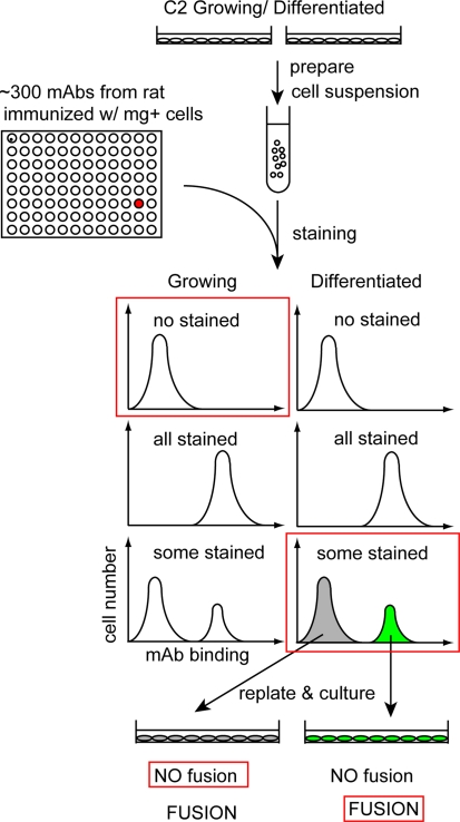
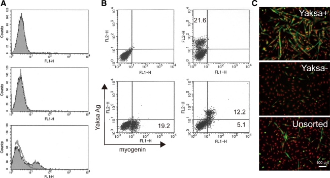
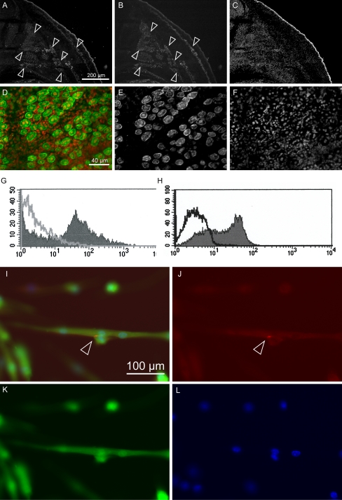
We established a novel monoclonal antibody, Yaksa that is specific to a subpopulation of myogenic cells. The Yaksa antigen is not expressed on the surface of growing myoblasts but only on a subpopulation of myogenin-positive myocytes. When Yaksa antigen-positive mononucleated cells were freshly prepared from a murine myogenic cell by a cell sorter, they fused with each other and formed multinucleated myotubes shortly after replating while Yaksa antigen-negative cells scarcely generated myotubes. These results suggest that Yaksa could segregate fusion-competent, mononucleated cells from fusion-incompetent cells during muscle differentiation. The Yaksa antigen was also expressed in developing muscle and regenerating muscle in vivo and it was localized at sites of cell-cell contact between mono-nucleated muscle cells and between mono-nucleated muscle cells and myotubes. Thus, Yaksa that marks prefusion myocytes before myotube formation can be a useful tool to elucidate the cellular and molecular mechanisms of myogenic cell fusion.


Figures

Fig. 1
Fig. 1
High correlation between GFP and myogenin expression in mgEGFP-C2. mgEGFP-C2 culture at 2 days after induction of differentiation (AD), and in 10%CS-DMEM (EH) was stained. A, E: phase contrast; B, F: anti-myogenin staining; C, G: GFP; D, H: DAPI staining
Fig. 2
Fig. 2
Screening procedure. Mononucleated cell suspension was prepared from either growing or differentiated C2, stained with mAbs generated from rat immunized with myogenin-positive cells, and sorted. Staining pattern was categorized to three groups: No stained, All stained, and Some stained. We selected a mAb which does not stain growing C2 but some C2 cells after differentiation (red boxed). Then if the cells reactive with a mAb form myotubes efficiently by a replating assay (red boxed), we assumed that the mAb reacted with prefusion myocytes. For details, see “Materials and methods”, and “Results”
Fig. 3
Fig. 3
Characterization of Yaksa. A FACScan analysis of Yaksa antigen on C2 cells. Upper growing, middle just before induction, lower 1.5 days after induction, dashed line control IgG2a. B Subpopulation of myogenin-positive cells express Yaksa antigen. FL1: myogenin stained with Alexa488, FL2; Yaksa antigen stained with PE. Thirty-six hours after induction, C2 cells were stained with control IgG (Upper Left), anti mg Ab (Lower Left), Yaksa (Upper Right), and anti mg Ab and Yaksa (Lower Right). C Yaksa-positive cells form multinucleated cells shortly after replating. Thirty-six hours after induction, C2 cells sorted with Yaksa were replated, cultured for 6 h, and then stained with anti-alpha actinin (green) and DAPI (red). Yaksa-positive cells form binuclear or multinucleated cells shortly after replating
Fig. 4
Fig. 4
Yaksa antigen was expressed in vivo. AC Transverse section of mouse embryo (E13.5) triple-stained with Yaksa (A), anti-Desmin (B) and DAPI (C). A dorsal quarter of embryo was shown. Arrow heads Desmin positive developing muscle. Signal in the peripheral of section is non-specific staining. DF Transverse section of regenerating TA muscle 4 days after CTX injection was stained with Yaksa (green, E) and DAPI (red, F). G FACScan analysis of single cells prepared from regenerating TA muscle 3 days after CTX injection. Cells were stained with Yaksa (solid) or IgG2a (line). H FACScan analysis of primary myoblasts prepared from gastrocnemius muscle. Cells were stained with Yaksa (solid) or IgG2a (line). IL Yaksa antigen is expressed at site of cell–cell contact. Cultures of pMB expressing GFP (green, K) were fixed and stained with biotinylated Yaksa & StAv-ALEXA594 (red, J) and Hoechst (blue, L). Arrowheads Accumulation of Yaksa antigen at sites of cell–cell contact

Similar articles

References

    1. Andres V, Walsh K. Myogenin expression, cell cycle withdrawal, and phenotypic differentiation are temporally separable events that precede cell fusion upon myogenesis. J Cell Biol. 1996;132:657–666. doi: 10.1083/jcb.132.4.657. - DOI - PMC - PubMed
    1. Blanco-Bose WE, Yao CC, Kramer RH, Blau HM. Purification of mouse primary myoblasts based on alpha 7 integrin expression. Exp Cell Res. 2001;265:212–220. doi: 10.1006/excr.2001.5191. - DOI - PubMed
    1. Buckingham M. Skeletal muscle formation in vertebrates. Curr Opin Genet Dev. 2001;11:440–448. doi: 10.1016/S0959-437X(00)00215-X. - DOI - PubMed
    1. Buckingham M. Myogenic progenitor cells and skeletal myogenesis in vertebrates. Curr Opin Genet Dev. 2006;16:525–532. doi: 10.1016/j.gde.2006.08.008. - DOI - PubMed
    1. Capkovic KL, Stevenson S, Johnson MC, Thelen JJ, Cornelison DD. Neural cell adhesion molecule (NCAM) marks adult myogenic cells committed to differentiation. Exp Cell Res. 2008;314:1553–1565. doi: 10.1016/j.yexcr.2008.01.021. - DOI - PMC - PubMed

Publication types