Review
. 2011 Nov;216(4):301-17.
doi: 10.1007/s00429-011-0322-0. Epub 2011 May 20.

Hippocampal pyramidal cells: the reemergence of cortical lamination

Affiliations
Review

Hippocampal pyramidal cells: the reemergence of cortical lamination

Lutz Slomianka et al. Brain Struct Funct. 2011 Nov.

Abstract

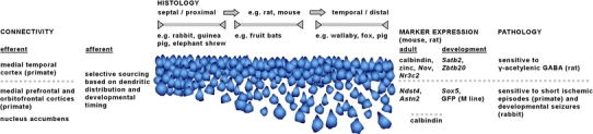
The increasing resolution of tract-tracing studies has led to the definition of segments along the transverse axis of the hippocampal pyramidal cell layer, which may represent functionally defined elements. This review will summarize evidence for a morphological and functional differentiation of pyramidal cells along the radial (deep to superficial) axis of the cell layer. In many species, deep and superficial sublayers can be identified histologically throughout large parts of the septotemporal extent of the hippocampus. Neurons in these sublayers are generated during different periods of development. During development, deep and superficial cells express genes (Sox5, SatB2) that also specify the phenotypes of superficial and deep cells in the neocortex. Deep and superficial cells differ neurochemically (e.g. calbindin and zinc) and in their adult gene expression patterns. These markers also distinguish sublayers in the septal hippocampus, where they are not readily apparent histologically in rat or mouse. Deep and superficial pyramidal cells differ in septal, striatal, and neocortical efferent connections. Distributions of deep and superficial pyramidal cell dendrites and studies in reeler or sparsely GFP-expressing mice indicate that this also applies to afferent pathways. Histological, neurochemical, and connective differences between deep and superficial neurons may correlate with (patho-) physiological phenomena specific to pyramidal cells at different radial locations. We feel that an appreciation of radial subdivisions in the pyramidal cell layer reminiscent of lamination in other cortical areas may be critical in the interpretation of studies of hippocampal anatomy and function.


Figures

Fig. 1
Fig. 1
Illustration of the nomenclature used in this review. The top left inset shows the hippocampus of one hemisphere from which non-hippocampal tissue had been removed and illustrates the terms “septal” and “temporal”. The approximately mid-septotemporal location of the main image of a horizontal section is indicated in the top left inset by a line. The main image illustrates the terms “proximal” and “distal”. The proximal border of CA1 (more correctly CA1/2) towards CA3 and between distal CA1 and the subiculum is marked by arrowheads. The top right inset shows the pyramidal cells marked in the main image and illustrates the use of the terms “superficial” and “deep”. Scalebar 100 μm
Fig. 2
Fig. 2
Nissl-stained CA1 pyramidal cell layer. Unless noted otherwise, images were taken at mid-proximodistal and mid-septotemporal locations. a Human, slight differences between superficial and deep CA1 can be seen in both cell density and staining characteristics of the pyramidal cells, b Marmoset monkey, c large-eared slit-faced bat, d little free-tailed bat, e Wahlberg’s epauletted fruit bat, f Parma wallaby, g common brush-tailed possum, h fox, i common mole rat, j eastern rock elephant shrew, k long-tailed chinchilla, l–n Wistar rat, l septal, m septal extreme and n temporal CA1, op C57BL/6 mouse, o septal and p temporal CA1. Scalebars al 50 μm, mp 25 μm
Fig. 3
Fig. 3
a Hippocampus of a mouse carrying the Reln orl reelin mutation. b Two distinct cell layers and deep cell clusters in the septal mid-proximodistal CA1 of a Reln orl mouse. c At more temporal levels packing densities and cell sizes in the two CA1 layers resemble those in normal laboratory mice. d CA1 pyramidal cells in Reln orl mouse retain their neurochemical identity with regard to calbindin, with only the younger, now deep pyramids showing moderate calbindin immunoreactivity. e The two CA1 layers of Reln orl mice do not show appreciable differences in their parvalbumin immunoreactivity. f Zbtb20 (green) in C57Bl/6 mouse superficial pyramidal cells and Sox5 (red) in deep pyramidal cells. g Scattered deep pyramidal cells in distal CA3 of the Chincilla do not receive mossy fiber input close to their soma. h Scattered deep pyramidal cells in distal CA3 of the fox. Scalebars a 0.5 mm, bh 50 μm. The original of f was kindly provided by Prof. Niels A. Jensen (Nielsen et al. , with permission of Oxford University Press)
Fig. 4
Fig. 4
Calbindin-immunoreactive (CaBP-ir) and green-fluorescent protein expressing pyramidal cells in a the septal CA1 and b the distal mid-septotemporal CA1 of thy1-GFP (M line) transgenic mice. c CaBP-ir CA1 pyramidal cells in septal CA1 of Wistar rat. Some of the voids left by unstained deep cells are marked with an asterisk. d Zinc-containing pyramidal cells in mid-septotemporal, mid-proximodistal CA1 of Wistar-Kyoto rat. e CaBP-ir CA1 pyramidal cells in temporal one-half of th fox CA1. Lightly CaBP-ir superficial cells are separated from a tier of strongly CaBP-ir, large deep pyramids by virtually unstained cells. f Cells in the septal three-quarters of the elephant shrew CA1 pyramidal cell layer (delimited by open arrows) are completely unstained. The filled arrow marks the boundary between stratum radiatum and stratum lacunosum-moleculare. g CaBP-ir deep CA1 pyramidal cells in the temporal CA1 of the elephant shrew. h CaBP-ir deep CA3 pyramidal cells in naked mole rats, mf: CaBP-ir mossy fibers. Scalebars a, c and d 20 μm; b, g and h 50 μm; e and f 100 μm
Fig. 5
Fig. 5
Mid-septotemporal hippocampus of Wistar Kyoto rats, stained for zinc-containing neurons and fluorescence of the retrograde tracer fluorogold. a In proximal CA1, predominantly deep zinc-negative pyramids label after an injection into the ipsilateral lateral septum. b After a contralateral fluorogold injection into the lateral septum, deep zinc-containing CA3 pyramids are not labeled, whereas c deep zinc-containing CA3 pyramids are labeled after an ipsilateral fluorogold injection into the lateral septum. d Only deep zinc-negative pyramidal cells in distal CA1 are retrogradely labeled after an ipsilateral fluorogold injection into the ventral striatum. Scalebars ac 50 μm; d 25 μm
Fig. 6
Fig. 6
Examples of particularly interesting histoarchitectural, connectional, neurochemical and pathological findings that distinguish superficial and deep pyramidal cells of CA1. The cell layer has been drawn to resemble the different appearances of the layer along both the septotemporal and proximodistal axis in mouse and rat. In other species much of CA1 is dominated by a histoarchitectural phenotype that encompasses only a narrow segment of the variations seen in rat and mouse

Similar articles

Cited by

References

    1. Abbie AA. The relations of the fascia dentata, hippocampus and neocortex, and the nature of the subiculum. J Comp Neurol. 1938;68:307–333. doi: 10.1002/cne.900680303. - DOI
    1. Altemus KL, Lavenex P, Ishizuka N, Amaral DG. Morphological characteristics and electrophysiological properties of CA1 pyramidal neurons in macaque monkeys. Neuroscience. 2005;136:741–756. doi: 10.1016/j.neuroscience.2005.07.001. - DOI - PubMed
    1. Altman J, Bayer SA. Prolonged sojourn of developing pyramidal cells in the intermediate zone of the hippocampus and their settling in the stratum pyramidale. J Comp Neurol. 1990;301:343–364. doi: 10.1002/cne.903010303. - DOI - PubMed
    1. Amaral DG. Emerging principles of intrinsic hippocampal organization. Curr Opin Neurobiol. 1993;3:225–229. doi: 10.1016/0959-4388(93)90214-J. - DOI - PubMed
    1. Amaral DG, Dolorfo C, Alvarez-Royo P. Organization of CA1 projections to the subiculum: a PHA-L analysis in the rat. Hippocampus. 1991;1:415–436. doi: 10.1002/hipo.450010410. - DOI - PubMed

Publication types

MeSH terms

LinkOut - more resources