. 2011 Nov;105(2):241-51.
doi: 10.1007/s11060-011-0604-7. Epub 2011 May 20.

Vorinostat modulates cell cycle regulatory proteins in glioma cells and human glioma slice cultures

Affiliations

Vorinostat modulates cell cycle regulatory proteins in glioma cells and human glioma slice cultures

Jihong Xu et al. J Neurooncol. 2011 Nov.

Abstract

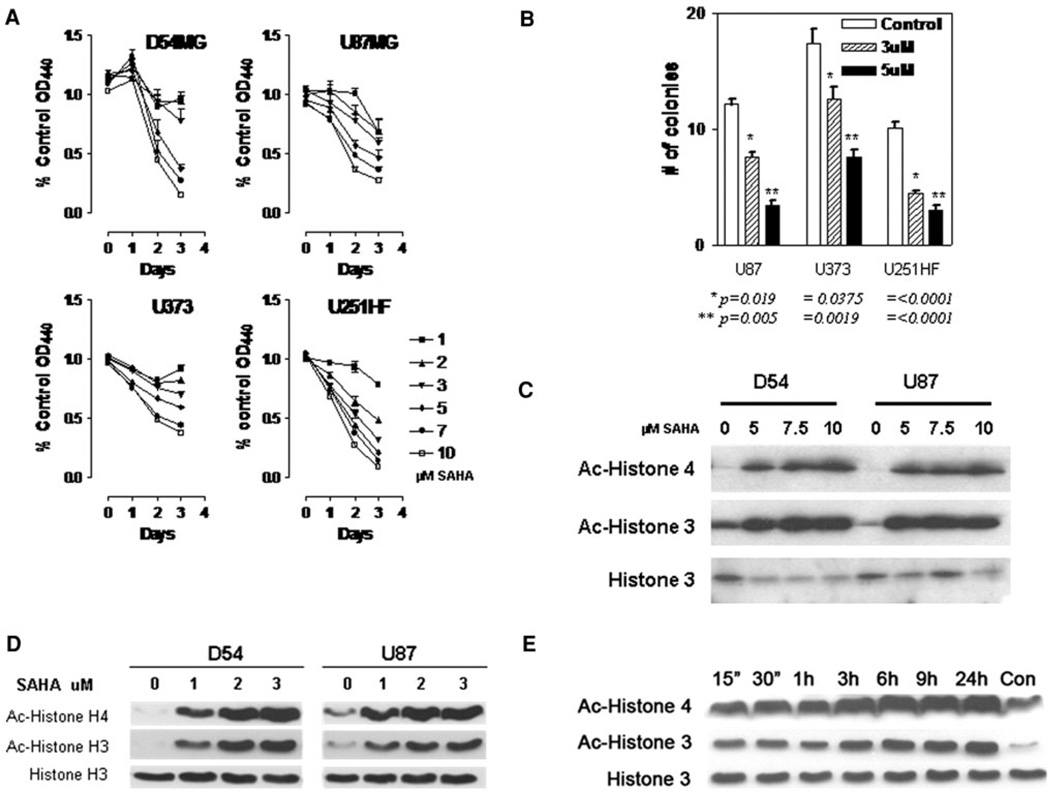
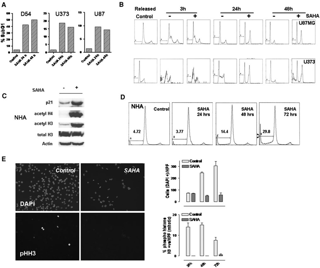
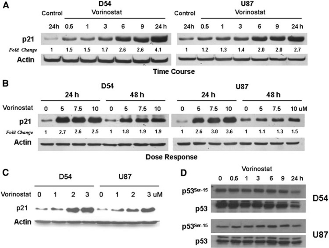
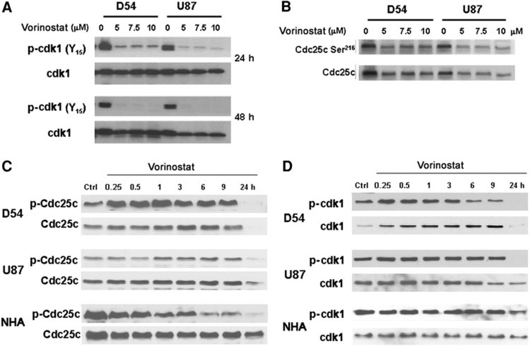
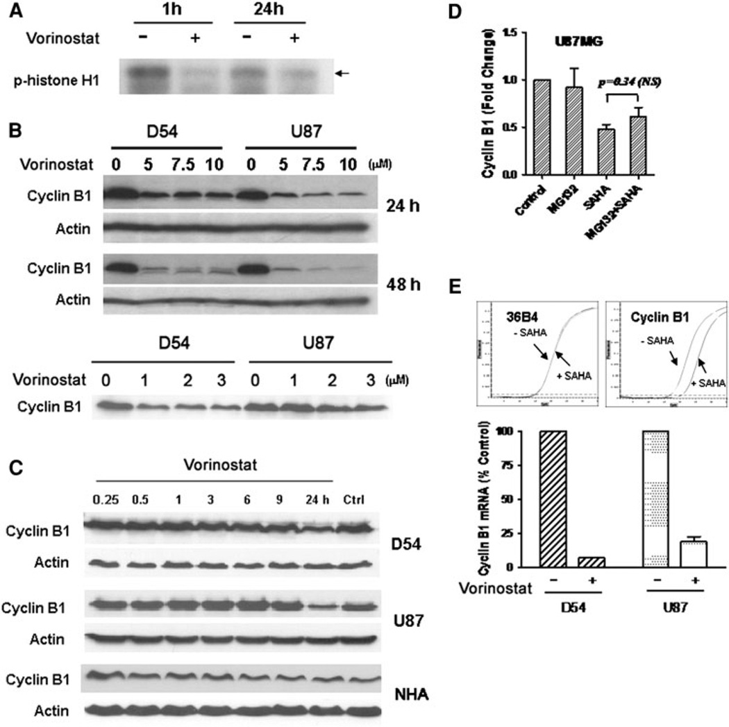
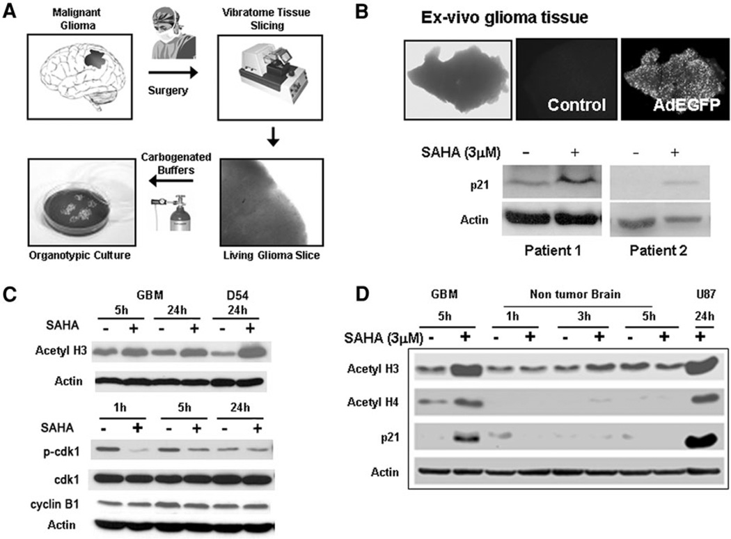
Chromatin modification through histone deacetylase inhibition has shown evidence of activity against malignancies. The mechanism of action of such agents are pleiotropic and potentially tumor specific. In this study, we studied the mechanisms of vorinostat-induced cellular effects in gliomas. The effects of vorinostat on proliferation, induction of apoptosis and cell cycle effects were studied in vitro (D54, U87 and U373 glioma cell lines). To gain additional insights into its effects on human gliomas, vorinostat-induced changes were examined ex vivo using a novel organotypic human glioma slice model. Vorinostat treatment resulted in increased p21 levels in all glioma cells tested in a p53 independent manner. In addition, cyclin B1 levels were transcriptionally downregulated and resulted in reduced kinase activity of the cyclin B1/cdk1 complex causing a G2 arrest. These effects were associated with a dose- and time-dependent inhibition of cellular proliferation and anchorage-independent growth in association with hyperacetylation of core histones and induction of apoptosis. Of particular significance, we demonstrate histone hyperacetylation and increased p21 levels in freshly resected human glioma specimens maintained as organotypic slice cultures and exposed to vorinostat similar to cell lines suggesting that human glioma can be targeted by this agent. Our data suggest that the effects of vorinostat are associated with modulation of cell cycle related proteins and activation of a G2 checkpoint along with induction of apoptosis. These effects are mediated by both transcriptional and post-translational mechanisms which provide potential options that can be exploited to develop new therapeutic approaches against gliomas.


Conflict of interest statement

Conflict of interest The authors declare no conflicts of interest related to this study.

Figures

Fig. 1
Fig. 1
a Glioma cells were exposed to vorinostat at various concentrations as indicated and the proliferation rate assessed by change in colorimetric values using a WST-1 assay, b Cells were plated in soft agar at a density of 10 cells/well in six well plates and anchorage independent growth determined by determining nuMber of colonies formed. Data shown represent colony counts averaged from five high powered fields (error bars = standard error of mean; statistical analysis performed between control and treatment indicated using a unpaired t-test with a two-tailed P value with significant values being ≤0.05). c D54 and U87 glioma cells were treated with higher concentrations (5–10 µM) of vorinostat for 24 h and acetyla-tion of histones H3 and H4 were determined by immunoblotting. d Effect of lower concentrations (1–3 µM) of vorinostat on histone acetylation was assessed by immunoblotting. e Time course of histone acetylation after treatment of D54 cells with 3 µM vorinostat (untreated control at 24 h is shown in rightmost lane)
Fig. 2
Fig. 2
a D54, U373 and U87 cells were treated with vorinostat (3 µM) for 24 and 48 h and analyzed for apoptotic cells by flowcytometric measurement of the sub G1 cell cycle fraction. b Cells were synchronized in the G1 phase cell cycle boundary by a thymidine/urea double block, treated with vorinostat (3 µM) and released. Cells in each condition were harvested at the periods indicated and analyzed by flowcytometry. Data shown are representative of two independent experiments. c Normal huMan astrocytes (NHA) treated with vorinostat (3 µM) or vehicle (DMSO) were assessed for changes in p21 levels, acetylation of histones and total histone levels after 48 h of exposure to the agent. d NHA, cultured in astrocyte basal medium, were treated with vorinostat (3 µM); flowcytometric analysis was performed to detect cell cycle changes and the sub-G1 fraction at the times indicated. e D54 cells were cultured on coverslips and exposed to vorinostat (3 µM) for the periods indicated; the cells were fixed and immunostained with DAPI (to label nuclei) and phospho histone H3 (for mitotic cells). Mitotic and total cells were counted after appropriate immunostaining. Data shown in graph are the mean counts of positive cells derived from two independent measurements
Fig. 3
Fig. 3
a D54 and U87 cells were exposed to vorinostat (3 µM) in a time-course experiment and harvested at the periods indicated. Levels of p21 were assessed by immunoblotting and actin levels were determined as a loading control. Fold change values (normalized to actin levels) compared to untreated control at 24 h are shown. b To determine the dose response relationship to vorinostat, cells were exposed to various higher concentrations (5–10 µM) of the agent and the levels of p21 determined by immunoblotting. c Changes in p21 levels were also assessed at lower concentrations (1–3 µM) of vorinostat with actin levels assessed as loading controls. d Phosphorylation status of p53 at Ser-15 was determined in comparison with total p53 levels at the periods indicated after exposure to vorinostat (3 µM)
Fig. 4
Fig. 4
a Changes in level of inhibitory phosphorylation of cdk1 after treatment with vorinostat was determined at 24 and 48 h (using a Tyr-15 residue phospho-specific antibody) along with total cdk1 as loading control. b Degree of inhibitory phosphorylation of cdc-25 at Ser-216 was assessed by immunoblotting in response to increasing doses of vorinostat. c Time course of changes in phospho-cdc25c levels after exposure to vorinostat (5 µM). d Time course of changes in levels of phospho-cdk1 and total cdk-1 were determined following treatment with vorinostat (5 µM)
Fig. 5
Fig. 5
a Cells treated with vorinostat (3 µM) for the periods indicated were assessed for kinase activity of the cdk1/cyclin B1 complex in an in vitro kinase assay using histone H1 as a substrate. The cell lysates were subsequently subject to immunoblotting using an anti phospho-histone H1 antibody, b Cyclin B1 levels were determined in D54 and U87 cells after treatment with various doses of vorinostat at 24 and 48 h (higher concentrations, upper panel; lower concentrations, lower panel) (note the actin control for the upper panel blot is shared by that for the p21 in Fig. 3c). c D54, U87 and NHA cells were treated with vorinostat (3 µM) and the time course of changes in cyclin B1 levels were assessed by immunoblotting. d U87MG cells were treated with vorinostat alone (3 µM), MG132 alone, or MG132 followed by vorinostat. Levels of cyclin B1 were determined by immunoblotting and the intensity of the bands were quantitated and expressed relative to levels in untreated control. Statistical analysis was performed between vorinostat alone or vorinostat plus MG132 using a unpaired t-test with a two-tailed P value; significant values are ≤ 0.05). e Levels of cyclin B1 transcript in D54 and U87 cells were determined by quantitative real-time PCR (upper panel) after treatment with vorinostat (3 µM) and with transcript levels of the ribosomal protein 36B4 as a control. Mean CT levels of cyclin B1 transcript (lower panel) are shown relative to levels of 36B4 transcript
Fig. 6
Fig. 6
a Generation of organotypic human glioma tissue slices: freshly resected tumor tissue was obtained from patients undergoing surgical removal of malignant gliomas and 300 urn thick slices were generated using a Vibratome. The slices were maintained in carbogenated ACSF and subsequently incubated at 37°C. b Tissue slices derived from freshly resected human glioblastoma specimens were incubated with vorinostat or vehicle control for 48 h and subsequently homogenized to generate protein lysates. Levels of p21 were determined by immunoblotting. Viability of the slices during the course of the experiment was assessed by simultaneously monitoring fluorescence signal from control slices transduced with an adenoviral construct expressing EGFP. c Glioma slices were treated with vorinostat (3 µM) or PBS for the periods indicated and the levels of histone acetylation, phospho-cdk1 and total cdk1, and cyclin B1 were determined by western blots. Actin levels were obtained as a loading control. d Tissue slices generated from a human glioblastoma specimen and from overlying non-tumor cortical brain tissue were treated with vorinostat (3 µM) and levels of acetyl histones and p21 determined in the lysates derived from the slice homogenates. Lysates from U87 cells treated with vorinostat (3 µM) were used as a control. Actin levels were determined both as a loading control and to determine the integrity of proteins in the tissue slices

References

    1. Holland EC. Gliomagenesis: genetic alterations and mouse models. Nat Rev. 2001;2:120–129. - PubMed
    1. Penas-Prado M, Gilbert MR. Molecularly targeted therapies for malignant gliomas: advances and challenges. Expert Rev Anticancer Ther. 2007;7:641–661. - PubMed
    1. Baylin SB. DNA methylation and gene silencing in cancer. Nat Clin Pract. 2005;2(Suppl 1):S4–S11. - PubMed
    1. Jones PA, Baylin SB. The epigenomics of cancer. Cell. 2007;128:683–692. - PMC - PubMed
    1. Amatya VJ, Naumann U, Weller M, Ohgaki H. TP53 promoter methylation in human gliomas. Acta Neuropathol. 2005;110:178–184. - PubMed

Publication types

MeSH terms