. 2011 Sep;112(9):2606-15.
doi: 10.1002/jcb.23187.

Tumor necrosis factor-α treatment of HepG2 cells mobilizes a cytoplasmic pool of ERp57/1,25D₃-MARRS to the nucleus

Affiliations

Tumor necrosis factor-α treatment of HepG2 cells mobilizes a cytoplasmic pool of ERp57/1,25D₃-MARRS to the nucleus

Brian J Grindel et al. J Cell Biochem. 2011 Sep.

Abstract

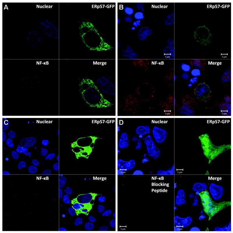
ERp57/PDIA3/1,25-MARRS has diverse functions and multiple cellular locations in various cell types. While classically described as an endoplasmic reticulum (ER) resident protein, ERp57 has a nuclear location sequence (NLS) and can enter the nucleus from the cytosol to alter transcription of target genes. Dysregulation and variable expression of ERp57 is associated with a variety of cancers including hepatocellular carcinoma (HCC). We investigated the dynamic mobility of ERp57 in an HCC cell line, HepG2, to better understand the movement and function of the non-ER resident pool of ERp57. Subcellular fractionation indicated ERp57 is highly expressed in the ER with a smaller cytoplasmic pool in HepG2 cells. Utilizing an ERp57 green fluorescent protein fusion construct created with and without a secretory signal sequence, we found that cytoplasmic ERp57 translocated to the nucleus within 15 min after tumor necrosis factor-α (TNF-α) treatment. Protein kinase C activators including 1,25-dihydroxyvitamin D(3) and phorbol myristate acetate did not trigger nuclear translocation of ERp57, indicating translocation is PKC independent. To determine if an interaction between the rel homology binding domain in ERp57 and the nuclear factor-κB subunit, p65, occurred after TNF-α treatment and could account for nuclear movement, co-immunoprecipitation was performed under control and conditions that stabilized labile disulfide bonds. No support for a functional interaction between p65 and ERp57 after TNF-α treatment was found in either case. Immunostaining for both ERp57-GFP and p65 after TNF-α treatment indicated that nuclear translocation of these two proteins occurs independently in HepG2 cells.


Figures

Figure 1
Figure 1. Proteins expressed off plasmid constructs
1A) The ER-mRFP construct encodes a monomeric red fluorescent protein (mRFP) preceded by a prolactin signal sequence and ending in a KDEL ER retention sequence for strong localization to the ER. The mRFP signal image below displays the ER-mRFP localization indicative of HepG2 cells. 1B) The ERp57-GFP construct encodes full length ERp57 with its natural signal sequence followed by a green fluorescent protein (GFP) and a KDEL ER retention sequence. The GFP signal image below shows the usual ERp57-GFP localization in HepG2 cells. 1C) The CytoERp57-GFP construct is the same as ERp57-GFP except it does not have a signal sequence. This lack of signal sequence results in ERp57 being diffusely expressed in the cytosol of HepG2 cells as seen in the bottom image.
Figure 2
Figure 2. ERp57 is highly expressed in HepG2 ER relative to cytoplasmic fractions
2A, B) Transiently transfected ERp57-GFP was highly co-localized with mRFP in the ER. Obvious green round structures were visible in some cells (white arrow 2A). Images at 100X objective, 2X zoom. 2C, D, E) Shown are the western blots for β-actin, BiP, and ERp57 of differentially centrifugated HepG2 cell fractions. The Methods section describes the differential centrifugation procedure and the text describes the results. H: homogenate; P6: resuspended pellet after 6,000×g spin; P105: resuspended pellet after 105,000×g spin (microsomal fraction); S105: supernatant after 105,000×g spin (cytoplasmic fraction).
Figure 3
Figure 3. ERp57-GFP translocates to the nucleus upon TNF-α treatment
HepG2 cells transiently transfected with ERp57-GFP were treated with vehicle (3 μl 0.1% BSA in PBS per 1000 μl SF DMEM) or 30 ng/ml TNF-α in SF DMEM for 1 hr or 15 min. The cells were stained for p65 (NF-κB, Red), and Draq5 nuclear stained (Blue, Nuclear). All are split channel images with the corresponding merge. A) 1 hr vehicle control treatment. B) 15 min TNF-α treatment. C) 1 hr TNF-α treatment. D) 15 min TNF-α treatment but p65 staining was blocked with a blocking peptide to show Ab specificity.
Figure 4
Figure 4. TNF-α enhances CytoERp57-GFP localization to the nucleus
HepG2 cells transfected with cytoERp57-GFP were treated with vehicle (3 μl 0.1% (w/v) BSA in PBS per 1000 μl SF DMEM) or 30 ng/ml TNF-α for 15 min. The cells were fixed, permeabilized, stained for p65 (NF-κB, Red), and Draq5 nuclear stained (Blue, Nuclear). All are split channel images with the corresponding merge. A) Vehicle treatment does not trigger nuclear translocation of p65, but some basal cytoERp57-GFP nuclear localization is seen. B) TNF-α treatment for 15 min increases the both the nuclear translocation of p65 and cytoERp57-GFP. However, p65 can have nuclear localization without cytoERp57-GFP (solid white arrow), and cytoERp57-GFP can have nuclear localization without p65 (dashed white arrow).
Figure 5
Figure 5. NF-κB and ERp57 do not associate in HepG2 cells after TNF-α treatment by Co-IP
Cell cultures were treated either with BSA vehicle control or 30ng/ml TNF-α for 15 min. The lysates were incubated either with p65 (NF-κB) or control IgG and western blotted for p65 and ERp57. A) Whole cell lysate B) incubated with p65 Ab C) incubated with control IgG. The arrows indicate the protein band for each western blot.

References

    1. Adikesavan AK, Unni E, Jaiswal AK. Overlapping signal sequences control nuclear localization and endoplasmic reticulum retention of GRP58. Biochem Biophys Res Commun. 2008;377:407–12. - PMC - PubMed
    1. Bourdi M, Demady D, Martin JL, Jabbour SK, Martin BM, George JW, Pohl LR. cDNA cloning and baculovirus expression of the human liver endoplasmic reticulum P58: characterization as a protein disulfide isomerase isoform, but not as a protease or a carnitine acyltransferase. Arch Biochem Biophys. 1995;323:397–403. - PubMed
    1. Chichiarelli S, Ferraro A, Altieri F, Eufemi M, Coppari S, Grillo C, Arcangeli V, Turano C. The stress protein ERp57/GRP58 binds specific DNA sequences in HeLa cells. J Cell Physiol. 2007;210:343–51. - PubMed
    1. Chignard N, Shang S, Wang H, Marrero J, Brechot C, Hanash S, Beretta L. Cleavage of endoplasmic reticulum proteins in hepatocellular carcinoma: Detection of generated fragments in patient sera. Gastroenterology. 2006;130:2010–22. - PubMed
    1. Cicchillitti L, Della Corte A, Di Michele M, Donati MB, Rotilio D, Scambia G. Characterisation of a multimeric protein complex associated with ERp57 within the nucleus in paclitaxel-sensitive and -resistant epithelial ovarian cancer cells: the involvement of specific conformational states of beta-actin. Int J Oncol. 2010;37:445–54. - PubMed

Publication types

MeSH terms

LinkOut - more resources