. 2011 Jun;39(3):725-32.
doi: 10.1042/BST0390725.

Crystallizing membrane proteins for structure-function studies using lipidic mesophases

Affiliations

Crystallizing membrane proteins for structure-function studies using lipidic mesophases

Martin Caffrey. Biochem Soc Trans. 2011 Jun.

Abstract

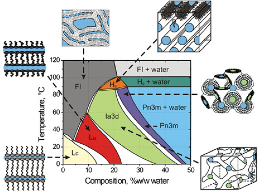
The lipidic cubic phase method for crystallizing membrane proteins has posted some high-profile successes recently. This is especially true in the area of G-protein-coupled receptors, with six new crystallographic structures emerging in the last 3½ years. Slowly, it is becoming an accepted method with a proven record and convincing generality. However, it is not a method that is used in every membrane structural biology laboratory and that is unfortunate. The reluctance in adopting it is attributable, in part, to the anticipated difficulties associated with handling the sticky viscous cubic mesophase in which crystals grow. Harvesting and collecting diffraction data with the mesophase-grown crystals is also viewed with some trepidation. It is acknowledged that there are challenges associated with the method. However, over the years, we have worked to make the method user-friendly. To this end, tools for handling the mesophase in the pico- to nano-litre volume range have been developed for efficient crystallization screening in manual and robotic modes. Glass crystallization plates have been built that provide unparalleled optical quality and sensitivity to nascent crystals. Lipid and precipitant screens have been implemented for a more rational approach to crystallogenesis, such that the method can now be applied to a wide variety of membrane protein types and sizes. In the present article, these assorted advances are outlined, along with a summary of the membrane proteins that have yielded to the method. The challenges that must be overcome to develop the method further are described.


Figures

Figure 1
Figure 1. Temperature–composition phase diagram of the mono-olein–water system determined under ‘conditions of use’ in the heating and cooling directions from 20°C
A cartoon representation of the various phase states is included in which coloured zones represent water. The liquid crystalline phases below ~17°C are metastable [13]. Abbreviations: FI, fluid isotropic phase; HII, inverted hexagonal phase; Lα, lamellar liquid crystalline phase; Lc, lamellar crystal phase. Figure reproduced from [24] with permission.
Figure 2
Figure 2. In meso crystallization model and crystals
(a) Schematic representation of the events proposed to take place during the crystallization of an integral membrane protein from the lipidic cubic mesophase. The process begins with the protein reconstituted into the highly curved bilayers of the bicontinuous cubic phase (bottom left quadrant). Added precipitants shift the equilibrium away from stability in the cubic membrane. This leads to phase separation, wherein protein molecules diffuse from the continuous bilayered reservoir of the cubic phase by way of a sheet-like or lamellar portal (upper left quadrant) to lock into the lattice of the advancing crystal face (upper right quadrant). Salt (positive and negative signs) facilitates crystallization, in part, by charge screening. Co-crystallization of the protein with native or added lipid (cholesterol) is shown in this illustration. As much as possible, the dimensions of the lipid (light yellow oval with tail), detergent (pink oval with tail), native membrane or added lipid (purple), protein (blue; β2-adrenergic receptor–T4 lysozyme; PDB code 2RH1), and bilayer and aqueous channels (dark blue) have been drawn to scale. The lipid bilayer is approximately 40 Å thick. Crystals of (b) BtuB, (c) bacteriorhodopsin and (d) light-harvesting complex II growing in meso. Reprinted, with permission, from the Annual Review of Biophysics [10a], Volume 38, © 2009 by Annual Reviews.
Figure 3
Figure 3
In meso crystal packing arrangement and molecular structures of membrane proteins. The packing arrangement is shown in columns 1–3 within each panel. Expanded views of individual proteins or oligomers are shown in columns 4 and 5. The view in columns 1 and 5 represents a projection along the stacking axis. The views in columns 2–4 are from within the plane of the stacked lamellae along the two other unit cell axes. In columns 1–3, black outlines are projections of the unit cell. Images in column 4 are from the Orientations of Proteins in Membranes (OPM) database (http://opm.phar.umich.edu/), in which the red and blue horizontal lines define the hydrophobic thickness of the protein. Proteins are identified by name, source organism, resolution and PDB code. Currently in meso records in the MPDB (http://www.mpdb.tcd.ie) number 44 for bacterial rhodopsins, six for the photosynthetic reaction centres, 13 for GPCR–T4 lysozyme chimaeras, three for gramicidin, two for β-barrels and one for light-harvesting complex II. The examples shown represent the highest resolution available for each of the six protein types [33] B. brevis, Bacillus brevis; H. salinarum, Halobacterium salinarum; E. coli, Escherichia coli; Rps. acidophila, Rhodopseudomonas acidophila; R. sphaeroides, Rhodobacter sphaeroides; H. sapiens, Homo sapiens.

References

    1. Rasmussen SGF, Choi H-J, Fung JJ, Pardon E, Casarosa P, Chae PS, DeVree BT, Rosenbaum DM, Thian FS, Kobilka TS, et al. Structure of a nanobody-stabilized active state of the β2 adrenoceptor. Nature. 2011;469:175–180. - PMC - PubMed
    1. Rosenbaum DM, Zhang C, Lyons JA, Holl R, Aragao D, Arlow DH, Rasmussen SGFR, Choi H-J, DeVree BT, Sunahara RK, et al. Structure and function of an irreversible agonist–β2 andrenoreceptor complex. Nature. 2011;469:236–240. - PMC - PubMed
    1. Warne A, Moukhametzianov R, Baker JG, Nehme R, Edwards PC, Leslie AGW, Schertler GFX, Tate CG. The structural basis for agonist and partial agonist action on a β1-adrenergic receptor. Nature. 2011;469:241–244. - PMC - PubMed
    1. Caffrey M. J.D. Bernal and the genesis of structural biology. J. Physics Conf. Ser. 2007;57:17–28.
    1. Caffrey M, Kinsella JE. Growth and acyltransferase activity of rabbit mammary gland during pregnancy and lactation. J. Lipid Res. 1977;18:44–52. - PubMed

Publication types

MeSH terms