

Inflammatory pseudotumor of the liver: a case report and review of the literature
- PMID: 21600001
- PMCID: PMC3123642
- DOI: 10.1186/1752-1947-5-196
Inflammatory pseudotumor of the liver: a case report and review of the literature
Abstract
Introduction: Inflammatory pseudotumor of the liver represents a fairly uncommon pathology. Although it is a benign tumor, the correct diagnosis can be missed.
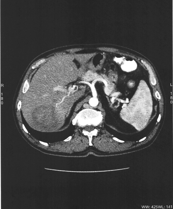
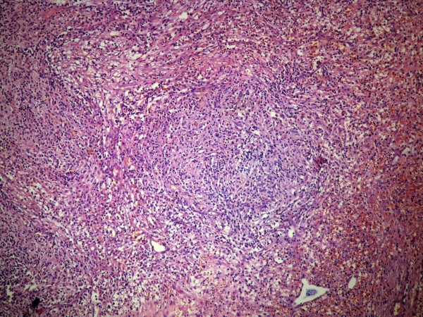
Case presentation: We report the case of a 55-year-old Caucasian man, who presented with a one-month history of abdominal pain and weight loss. He was diagnosed with a primary liver tumor by computed tomography and magnetic resonance imaging. Alpha-fetoprotein levels ranged within normal limits. A right posterior sectorectomy was performed. Histopathology revealed an inflammatory pseudotumor of the liver. Our patient remains in good condition one year later.
Conclusion: Although inflammatory pseudotumor of the liver is usually a benign process, controversy regarding its management still exists. With this case report we review the existing literature and consider hepatectomy as a safe treatment approach.
Figures

References
-
- Fukuya T, Honda H, Matsumata T, Kawanami T, Shimoda Y, Muranaka T, Hayashi T, Maeda T, Sakai H, Masuda K. Diagnosis of inflammatory pseudotumour of the liver: value of CT. AJR Am J Roentgenol. 1994;163(5):1087–1091. - PubMed
-
- Someren A. Inflammatory pseudotumour of liver with occlusive phlebitis: report of a case in child and review of the literature. Am J Clin Pathol. 1978;69(2):176–181. - PubMed
-
- Goldsmith PJ, Loganathan A, Jacob M, Ahmad N, Toogood GJ, Lodge JPA, Prasad KR. Inflammatory pseudotumours of the liver: a spectrum of presentation and management options. Eur J Surg Oncol. 2009;35(12):1295–1298. - PubMed
-
- Torzilli G, Inoue K, Midorikawa Y, Hui AM, Takayama T, Makuuchi M. Inflammatory pseudotumours of the liver: prevalence and clinical impact in surgical patients. Hepatogastroenterology. 2001;48(40):1118–1123. - PubMed
LinkOut - more resources
Full Text Sources
