

Iron, zinc and copper in the Alzheimer's disease brain: a quantitative meta-analysis. Some insight on the influence of citation bias on scientific opinion
- PMID: 21600264
- PMCID: PMC3134620
- DOI: 10.1016/j.pneurobio.2011.05.001
Iron, zinc and copper in the Alzheimer's disease brain: a quantitative meta-analysis. Some insight on the influence of citation bias on scientific opinion
Abstract
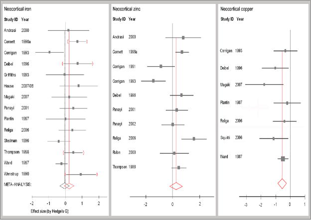
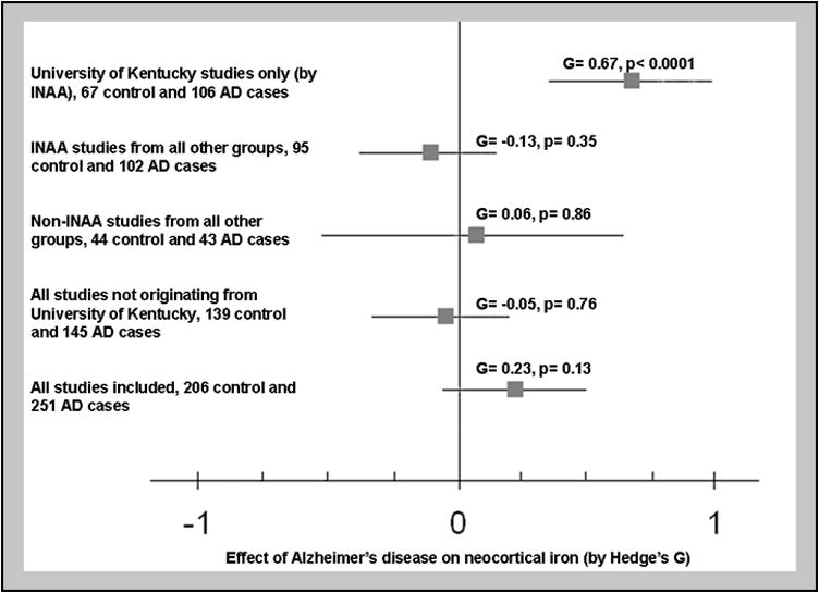
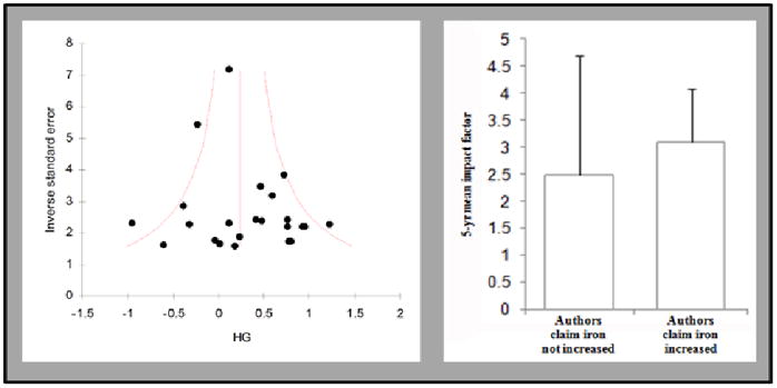
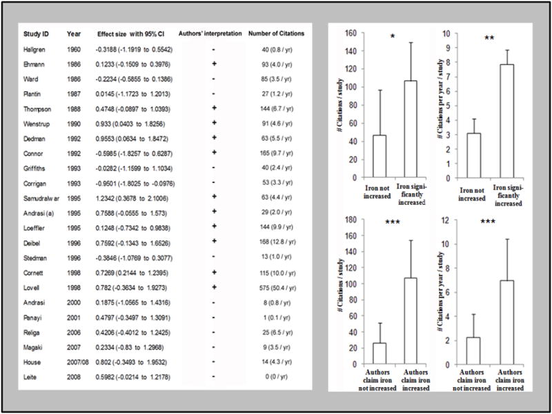
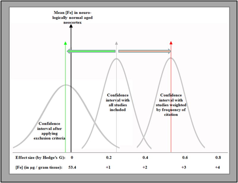
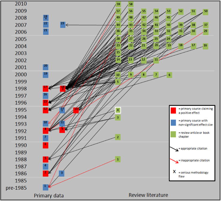
Dysfunctional homeostasis of transition metals is believed to play a role in the pathogenesis of Alzheimer's disease (AD). Although questioned by some, brain copper, zinc, and particularly iron overload are widely accepted features of AD which have led to the hypothesis that oxidative stress generated from aberrant homeostasis of these transition metals might be a pathogenic mechanism behind AD. This meta-analysis compiled and critically assessed available quantitative data on brain iron, zinc and copper levels in AD patients compared to aged controls. The results were very heterogeneous. A series of heavily cited articles from one laboratory reported a large increase in iron in AD neocortex compared to age-matched controls (p<0.0001) while seven laboratories failed to reproduce these findings reporting no significant difference between the groups (p=0.76). A more than three-fold citation bias was found to favor outlier studies reporting increases in iron and this bias was particularly prominent among narrative review articles. Additionally, while zinc was not significantly changed in the neocortex (p=0.29), copper was significantly depleted in AD (p=0.0003). In light of these findings, it will be important to re-evaluate the hypothesis that transition metal overload accounts for oxidative injury noted in AD.
Copyright © 2011 Elsevier Ltd. All rights reserved.
Figures
References
6. Annotated references
-
- Andrasi E, Farkas E, Gawlik D, Rosick U, Bratter P. Brain iron and zinc contents of German patients with Alzheimer’s disease. Journal of Alzheimer’s Disease. 2000;2:17C–26. INCLUDED: Iron and zinc were measured by INAA for hippocampus, frontal, parietal, occipital lobes, thalamus, caudate putamen and globus pallidus. Dry weight values were reported without dry to wet weight ratios; standard conversion ratios were applied. Results are from Eotvos University. - PubMed
-
- Andrasi E, Farkas E, Scheibler H, Reffy A, Bezur L. Al, Zn, Cu, Mn and Fe levels in brain in Alzheimer’s disease. Archives of Gerontology and Geriatrics. 1995;21:89–97. EXCLUDED for formalin fixation: iron, zinc and copper were measured from ten brain regions by INAA and ICP-AES. - PubMed
-
- Collingwood J, Dobson J. In situ characterization and mapping of iron compounds in Alzheimer’s disease tissue. Journal of Alzheimer’s Disease. 2005;7:267–72. EXCLUDED tissue fixation and semi-quantitative analysis: iron was measured by x-ray fluorescence in the frontal cortex of a single Alzheimer’s case. - PubMed
-
- Connor J, Snyder B, Beard J, Fine R, Mufson E. Regional distribution of iron and iron-regulatory proteins in the brain in aging and Alzheimer’s disease. Journal of Neuroscience Research. 1992;31:327–335. EXCLUDED for semi-quantitative methods (iron concentration normalized to protein concentration, not tissue weight): iron was measured by colorimetric assay in three brain regions. - PubMed
-
- Cornett C, Markesbery W, Ehmann W. Imbalances of trace elements related to oxidative damage in Alzheimer’s disease brain. Neurotoxicology. 1998a;19:339–45. INCLUDED: Iron and zinc were measured by INAA in hippocampus, amygdala, frontal, parietal and temporal lobes and cerebellum. Results are from the University of Kentucky. - PubMed
7. General references
-
- Amit T, Avramovich-Tirosh Y, Youdim M, Mandel S. Targeting multiple Alzheimer’s disease etiologies with multimodal neuroprotective and neurorestorative iron chelators. FASEB J. 2008;22:1296–305. - PubMed
-
- Atamna H. Amino acids variation in amyloid-beta peptides, mitochondrial dysfunction and new therapies for Alzheimer’s disease. J Bioenerg Biomembr. 2009;41:457–64. - PubMed
-
- Benzi G, Moretti A. Are reactive oxygen species involved in Alzheimer’s disease. Neurobiol Aging. 1995;16:661–74. - PubMed
-
- Brewer G. Iron and copper toxicity in diseases of aging, particularly atherosclerosis and Alzheimer’s disease. Exp Biol Med. 2007;232l:323–35. - PubMed
Publication types
MeSH terms
Substances
Grants and funding
LinkOut - more resources
Full Text Sources
Other Literature Sources
Medical
