. 2011 Sep;126(3-5):78-86.
doi: 10.1016/j.jsbmb.2011.05.001. Epub 2011 May 10.

The effect of tamoxifen and raloxifene on estrogen metabolism and endometrial cancer risk

Affiliations

The effect of tamoxifen and raloxifene on estrogen metabolism and endometrial cancer risk

Marian Y Williams-Brown et al. J Steroid Biochem Mol Biol. 2011 Sep.

Abstract

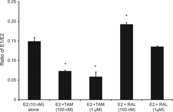
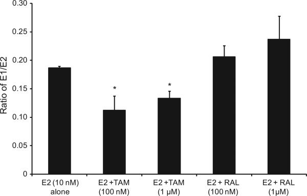
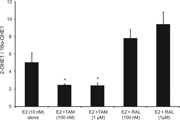
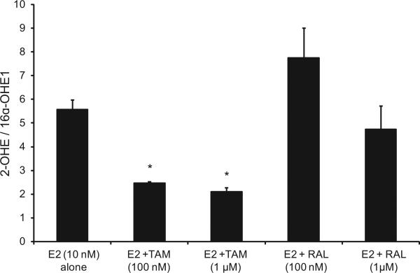
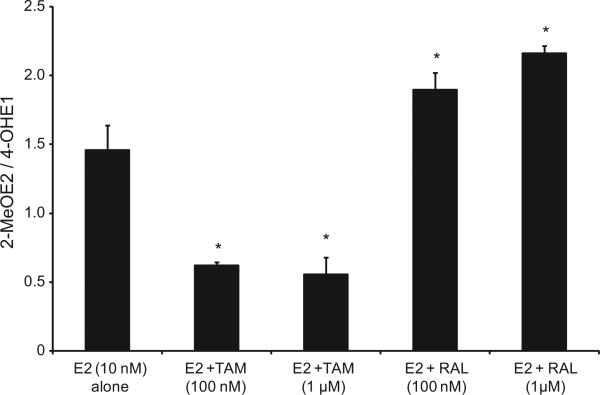
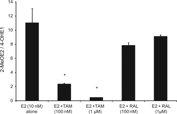
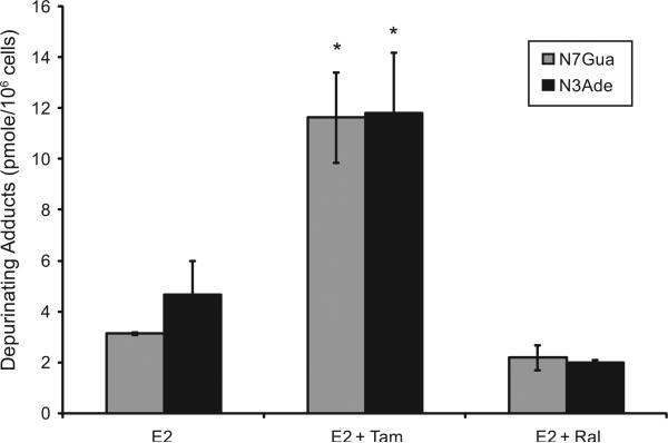
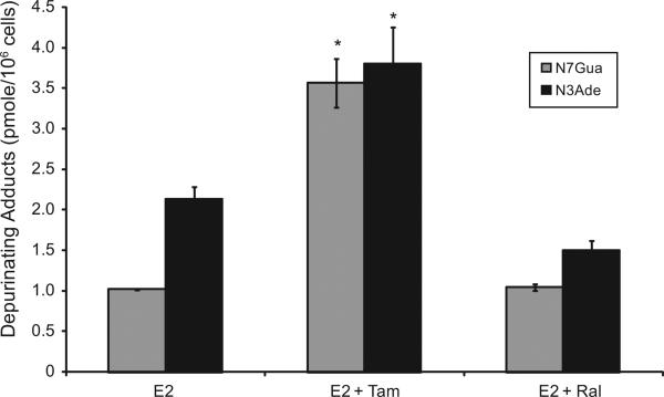
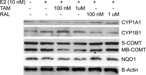
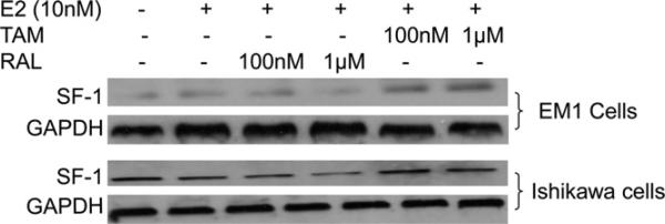
Selective estrogen receptor modulators (SERMs) demonstrate differential endometrial cancer (EC) risk. While tamoxifen (TAM) use increases the risk of endometrial hyperplasia and malignancy, raloxifene (RAL) has neutral effects on the uterus. How TAM increases the risk of EC and why TAM and RAL differentially modulate the risk for EC, however, remain elusive. Here, we tested the hypothesis that TAM increases the risk for EC, at least in part, by enhancing the local estrogen biosynthesis and directing estrogen metabolism towards the formation of genotoxic and hormonally active estrogen metabolites. In addition, the differential effects of TAM and RAL in EC risk are attributed to their differential effect on estrogen metabolism/metabolites. The endometrial cancer cell line (Ishikawa cells) and the nonmalignant immortalized human endometrial glandular cell line (EM1) were used for the study. The profile of estrogen/estrogen metabolites (EM), depurinating estrogen-DNA adducts, and the expression of estrogen-metabolizing enzymes in cells treated with 17β-estradiol (E2) alone or in combination with TAM or RAL were investigated using high performance liquid chromatography-electrospray ionization-tandem mass spectrometry (HPLC-ESI-MS(2)), ultraperformance liquid chromatography/tandem mass spectrometry (UPLC-MS/MS), and Western blot analysis, respectively. TAM significantly increased the total EM and enhanced the formation of hormonally active and carcinogenic estrogen metabolites, 4-hydroxestrone (4-OHE1) and 16α-hydroxyestrone, with concomitant reduction in the formation of antiestrogenic and anticarcinogenic 2-hydroxyestradiol and 2-methoxyestradiol. Furthermore, TAM increased the formation of depurinating estrogen-DNA adducts 4-OHE1 [2]-1-N7Guanine and 4-OHE1 [2]-1-N3 Adenine. TAM-induced alteration in EM and depurinating DNA adduct formation is associated with altered expression of estrogen metabolizing enzymes CYP1A1, CYP1B1, COMT, NQO1, and SF-1 as revealed by Western blot analysis. In contrast to TAM, RAL has minimal effect on EM, estrogen-DNA adduct formation, or estrogen-metabolizing enzymes expression. These data show that TAM perturbs the balance of estrogen-metabolizing enzymes and alters the disposition of estrogen metabolites, which can explain, at least in part, the mechanism for TAM-induced EC. These results also implicate the differential effect of TAM and RAL on estrogen metabolism/metabolites as a potential mechanism for their disparate effects on the endometrium.


Figures

Fig. 1
Fig. 1
The E1/E2 ratio was significantly lower compared with cells treated with E2 alone in EM1 cells treated with E2 plus TAM. The ratio of E1/E2 ratio in culture media from EM1 cells treated with E2 10 nM alone or with various concentrations of TAM or RAL. Cells were grown in serum-free media and treated for 48 h. Media were collected to quantify the E1/E2 ratio by HPLC–electrospray ionization-tandem mass spectrometry. Data were normalized against protein concentration. *Significant (P < 0.05) difference compared with treatment with E2 alone. Values represent the mean; bars represent the standard error of the mean.
Fig. 2
Fig. 2
The E1/E2 ratio was significantly lower compared with cells treated with E2 alone in Ishikawa cells treated with E2 plus TAM. The ratio of E1/E2 ratio in culture media from Ishikawa cells treated with E2 10 nM alone or with various concentrations of TAM or RAL as previously described. *Significant (P < 0.05) difference compared with treatment with E2 alone. Values represent the mean; bars represent the standard error of the mean.
Fig. 3
Fig. 3
Treatment with E2 and TAM significantly decreased the 2-OHE1/16α-OHE1 ratio compared with the treatment with E2 alone in EM1 cells; E2 and RAL increased this ratio. The ratio of 2-OHE1/16α- OHE1 ratio in culture media from EM1 cells treated with E2 10 nM alone or with various concentrations of TAM or RAL as previously described. *Significant (P < 0.05) difference compared with treatment with E2 alone. Values represent the mean; bars represent the standard error of the mean.
Fig. 4
Fig. 4
Treatment with E2 and TAM significantly decreased the 2-OHE1/16α-OHE1 ratio compared with the treatment with E2 alone in Ishikawa cells; RAL increased this ratio. The ratio of 2-OHE1/16α-OHE1 ratio in culture media from Ishikawa cells treated with E2 10 nM alone or with various concentrations of TAM or RAL as previously described. *Significant (P < 0.05) difference compared with treatment with E2 alone. Values represent the mean; bars represent the standard error of the mean.
Fig. 5
Fig. 5
Treatment with TAM significantly decreased the 2-MeOE2/4-OHE1 ratio in EM1 cells; treatment with RAL significantly increased this ratio. The ratio of 2ME/4-OHE1 ratio in culture media from EM1 cells treated with E2 10 nM alone or with various concentrations of TAM or RAL as previously described. *Significant (P < 0.05) difference compared with treatment with E2 alone. Values represent the mean; bars represent the standard error of the mean.
Fig. 6
Fig. 6
Treatment with TAM significantly decreased the 2-MeOE2/4-OHE1 ratio in Ishikawa cells. The ratio of 2ME/4-OHE1 ratio in culture media from Ishikawa cells treated with E2 10 nM alone or with various concentrations of TAM or RAL as previously described. *Significant (P < 0.05) difference compared with treatment with E2 alone. Values represent the mean; bars represent the standard error of the mean.
Fig. 7
Fig. 7
TAM significantly increased the levels of depurinating DNA adducts in EM1 cells. The levels of DNA adducts (4-OHE1 [2]-1-N7Guanine and 4-OHE1 [2]-1-N3 Adenine), were determined in culture media from EM-1 endometrial cells treated with either E2 (10 nM) alone or in combination with either TAM (1 μM) or RAL (1 μM) by ultraperformance liquid chromatography/tandem mass spectrometry (UPLC–MS/MS).
Fig. 8
Fig. 8
TAM significantly increased the levels of depurinating DNA adducts in Ishikawa cells. The levels of DNA adducts (4-OHE1 [2]-1-N7Guanine and 4-OHE1 [2]-1-N3 Adenine) were determined in culture media from Ishikawa endometrial cells treated with either E2 (10 nM) alone or in combination with either TAM (1 μM) or RAL (1 μM) by ultraperformance liquid chromatography/tandem mass spectrometry (UPLC–MS/MS).
Fig. 9
Fig. 9
Expression of CYP1A1, CYP1B1, S-COMT, MB-COMT, NQO1, and β-actin in EM1 cells. Cells were treated with E2 alone or in combination with various concentrations of TAM or RAL. Total cell lysates, nuclear fraction, and cytoplasmic fraction were prepared as described in Section 2. CYP1A1, CYP1B1, S-COMT, MB-COMT, NQO1, and β-actin expression were determined using Western blot analysis. The level of CYP1B1 was increased by E2 and further increased by addition of TAM. The expression of CYP1A1 was decreased by E2 and further decreased by addition of TAM. E2 alone or in combination with TAM resulted in a marked reduction in COMT and NQO1 expression compared with the control.
Fig. 10
Fig. 10
Expression of SF-1 and GAPDH in EM1 cells and Ishikawa cells. Cells were treated with E2 alone or in combination with various concentrations of TAM or RAL. Total cell lysates, nuclear fraction, and cytoplasmic fraction were prepared as described in the materials and methods section. SF-1 and GAPDH expression was determined using Western blot analysis. The level of SF-1 was increased by addition of TAM in EM1 and Ishikawa cells.

References

    1. Cohen I. Endometrial pathologies associated with postmenopausal tamoxifen treatment. Gynecol. Oncol. 2004;94:256–266. - PubMed
    1. Neven P, Vergote I. Controversies regarding tamoxifen and uterine carcinoma. Curr. Opin. Obstet. Gynecol. 1998;10:9–14. - PubMed
    1. Slomovitz BM, Sun CC, Ramirez PT, Bodurka DC, Diaz P, Lu KH. Does tamoxifen use affect prognosis in breast cancer patients who develop endome-trial cancer? Obstet. Gynecol. 2004;104:255–260. - PubMed
    1. DeMichele A, Troxel AB, Berlin JA, Weber AL, Bunin GR, Turzo E, Schinnar R, Burgh D, Berlin M, Rubin SC, Rebbeck TR, Strom BL. Impact of raloxifene or tamoxifen use on endometrial cancer risk: a population-based case–control study. J. Clin. Oncol. 2008;26:4151–4159. - PMC - PubMed
    1. Hemminki K, Rajaniemi H, Lindahl B, Moberger B. Tamoxifen-induced DNA adducts in endometrial samples from breast cancer patients. Cancer Res. 1996;56:4374–4377. - PubMed

Publication types

MeSH terms