. 2011 Jul 1;410(1):131-45.
doi: 10.1016/j.jmb.2011.04.077. Epub 2011 May 13.

Regulating SR protein phosphorylation through regions outside the kinase domain of SRPK1

Affiliations

Regulating SR protein phosphorylation through regions outside the kinase domain of SRPK1

Ryan M Plocinik et al. J Mol Biol. .

Erratum in

  • J Mol Biol. 2011 Aug 12;411(2):511. Whitehouse, Jennifer [corrected to Whitesides, Jennifer]

Abstract

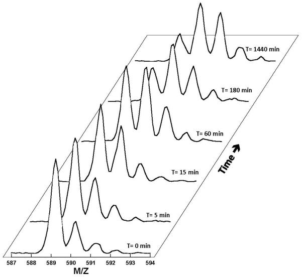
SR proteins (splicing factors containing arginine-serine repeats) are essential splicing factors whose phosphorylation by the SR-specific protein kinase (SRPK) family regulates nuclear localization and mRNA processing activity. In addition to an N-terminal extension with unknown function, SRPKs contain a large, nonhomologous spacer insert domain (SID) that bifurcates the kinase domain and anchors the kinase in the cytoplasm through interactions with chaperones. While structures for the kinase domain are now available, constructs that include regions outside this domain have been resistant to crystallographic elucidation. To investigate the conformation of the full-length kinase and the functional role of noncatalytic regions, we performed hydrogen-deuterium exchange and steady-state kinetic experiments on SRPK1. Unlike the kinase core, the large SID lacks stable, hydrogen-bonded structure and may provide an intrinsically disordered region for chaperone interactions. Conversely, the N-terminus, which positively regulates SR protein binding, adopts a stable structure when the insert domain is present and stabilizes a docking groove in the large lobe of the kinase domain. The N-terminus and SID equally enhance SR protein turnover by altering the stability of several catalytic loop segments. These studies reveal that SRPK1 uses an N-terminal extension and a large, intrinsically disordered region juxtaposed to a stable structure to facilitate high-affinity SR protein interactions and phosphorylation rates.


Figures

Figure 1
Figure 1. Effects of N-terminus & SID on Phosphorylation of SRSF1
A) SRPK1 and SRSF1 Constructs. B) Steady-State Kinetics. Initial velocities are normalized to Vmax and plotted as a function of total SRSF1 for SRPK1 (●), SRPK1(ΔN) (■), and SRPK1(ΔS) (▲). Kinetic parameters derived from hyperbolic fitting are displayed in Table 1. C) Competition experiments. Relative initial velocities for the phosphorylation of 50 nM SR(ΔRRM2) is monitored as a function of increasing concentrations of SRSF1 for SRPK1, SRPK1(ΔN), and SRPK1(ΔS). Proteins are labeled as in panel B and KI values are displayed in Table 1. D) Single turnover experiments. SRSF1 (100 nM) is preequilbrated with 2 μM of SRPK1, SRPK1(ΔN), and SRPK1(ΔS) and the reaction is started with 32P-ATP. Proteins are labeled as in panel B. The progress curves for SRPK1 are fit to a double exponential with rate constants and amplitudes of 6 and 0.2 min−1 and 12 and 3 sites. The progress curves for SRPK1(ΔN) and SRPK1(ΔS) are fit to single exponential functions with a common rate constant of 0.5 min−1 and amplitudes of 14 and 13 sites. E) Directionality Experiments. Complexes of enzymes (2 mM) and clASF(214) (200 nM) are allowed to react with varying concentrations of 32P-ATP and then cleaved with LysC to obtain N- and C-terminal fragments on SDS-PAGE. Relative 32P in both bands (N/C) are plotted as a function of total phosphoryl content. Symbols are the same as in panels B-D.
Figure 2
Figure 2. Binding of Fluorescently-Labeled Arg-Ser Peptides to SRPK1 and Deletion Mutants
(A-B) Fluorescence emission spectra of 10 nM fl(RS)8 (A) and fl(RS)16 (B) in the absence (black) and presence of 25 nM (red), 50 nM (green) and 75 nM (blue) SRPK1. (C-D) Plots of changes in fluorescence at 520 nM (normalized to total fluorescence change, ΔFmax) as a function varying SRPK1 (●), SRPK1(ΔN) (■), and SRPK1(ΔS) (▲). The Kd values are displayed in Table 2.
Figure 3
Figure 3. Mass Spectral Shift Upon Solvent Deuterium Incorporation
Mass spectra of a peptide probe (residues 143-147) from SRPK1 are recorded as a function of time.
Figure 4
Figure 4. Time-Dependent Deuterium Incorporation Into SRPK1 Probes
Deuteration levels of probes as a function of time are colored coded and assigned to the primary structure in SRPK1. Regions of secondary structure known from crystallographic data are assigned above the residues. Regions not defined (N-terminus & SID) are displayed as dotted brackets above the primary structure whereas those defined by X-ray crystallographic data (kinase 1 & 2) are displayed with solid brackets above the primary structure.
Figure 5
Figure 5. Effects of N-Terminal and SID Deletion on Time-Dependent Deuterium Incorporation Into SRPK1 Probes
Solvent deuterium incorporation as a function time for several probes are plotted for SRPK1 (red) SRPK1(ΔN) (blue) and SRPK1(ΔS) (green). The probes are defined by residues 76-91 (A), 109-122 (B), 151-164 (C), 202-220 (D), 512-523 (E), 569-590 (F), 591-608 (G), and 635-647 (H). These probes encompass portions of the following structural elements: glycine-rich loop (A), helix αC (B), β4-5 (C), catalytic loop (D), activation loop (E), helix αG (F), the MAP kinase insert (G) and helix αI (H).
Figure 6
Figure 6. Probes Affected by Deletion Mapped to the SRPK1 X-Ray Core Structure
Probes showing faster H-D exchange in SRPK1(ΔN) or SRPK1(ΔS) relative to SRPK1 are colored red, probes showing slower exchange are colored blue and those that are unaffected by deletion are colored yellow. Regions lacking high quality overlapping probes are gray and regions present in the X-ray structure but not in SRPK1(ΔS) and SRPK1(ΔN) are black. Critical structural elements in the kinase core are labeled. The space-filling models are shown in the same orientation as the ribbon diagram and then rotated by 180° around the y-axis.
Figure 7
Figure 7. Molecular Connectivities in the N-Terminus
A) Steady-State Kinetic Effects of Charge-to-Ala Mutations. Charged residues in two regions of the N-terminus defined by available H-D exchange probes (boxes) were mutated to alanine to generate 6 mutant kinases (M1-6). The steady-state kinetic parameters were measured and plotted as initial velocity (normalized to the total enzyme concentration) versus total SRSF1 concentrations. The steady-state kinetic parameters are displayed in the bar graph relative to the wild-type parameters for kcat and SRSF1 Km values. For comparison, the data for SRPK1(ΔN) are also displayed in the bar graph. B) Effects of the SID on H-D exchange in the N-Terminus. Deuteration levels of probes in the N-terminus of SRPK1(ΔS) are compared to the wild-type enzyme. Color coding and time parameters are the same as in Fig. 4.

References

    1. Jurica MS, Moore MJ. Pre-mRNA splicing: awash in a sea of proteins. Mol Cell. 2003;12:5–14. - PubMed
    1. Long JC, Caceres JF. The SR protein family of splicing factors: master regulators of gene expression. Biochem J. 2009;417:15–27. - PubMed
    1. Ghosh G, Adams JA. Phosphorylation mechanism and structure of serine-arginine protein kinases. FEBS J. 278:587–97. - PMC - PubMed
    1. Lai MC, Lin RI, Tarn WY. Transportin-SR2 mediates nuclear import of phosphorylated SR proteins. Proc Natl Acad Sci U S A. 2001;98:10154–9. - PMC - PubMed
    1. Hamelberg D, Shen T, McCammon JA. A proposed signaling motif for nuclear import in mRNA processing via the formation of arginine claw. Proc Natl Acad Sci U S A. 2007;104:14947–51. - PMC - PubMed

Publication types

MeSH terms