Review
. 2011 Jul 15;91(2):279-88.
doi: 10.1093/cvr/cvr098. Epub 2011 May 19.

Zebrafish as a model to study cardiac development and human cardiac disease

Affiliations
Review

Zebrafish as a model to study cardiac development and human cardiac disease

Jeroen Bakkers. Cardiovasc Res. .

Abstract

Over the last decade, the zebrafish has entered the field of cardiovascular research as a new model organism. This is largely due to a number of highly successful small- and large-scale forward genetic screens, which have led to the identification of zebrafish mutants with cardiovascular defects. Genetic mapping and identification of the affected genes have resulted in novel insights into the molecular regulation of vertebrate cardiac development. More recently, the zebrafish has become an attractive model to study the effect of genetic variations identified in patients with cardiovascular defects by candidate gene or whole-genome-association studies. Thanks to an almost entirely sequenced genome and high conservation of gene function compared with humans, the zebrafish has proved highly informative to express and study human disease-related gene variants, providing novel insights into human cardiovascular disease mechanisms, and highlighting the suitability of the zebrafish as an excellent model to study human cardiovascular diseases. In this review, I discuss recent discoveries in the field of cardiac development and specific cases in which the zebrafish has been used to model human congenital and acquired cardiac diseases.


Figures

Figure 1
Figure 1
Stages of cardiac development. (A) At 5 h post fertilization (hpf) the blastula (white) covers approximately 50% of the large yolk cell (yellow). At this stage, cardiac progenitor cells are located bilaterally in the lateral marginal zone. Atrial progenitor cells (pink) are located more ventrally than the ventricle progenitor cells (light blue). During gastrulation, the cardiac progenitor cells move dorsally towards the mid-line to end up in the anterior later plate mesoderm (ALPM). Cardiogenic differentiation is initiated in the future ventricle myocardial cells by the expression of cardiac myosins (purple) at the 12-somite stage. During mid- and late-somite stages, the myocardial tissue expands by continuous cardiogenic differentiation into more lateral regions of the ALPM by the cardiogenic differentiation of future atrial myocytes (orange; venous differentiation). Whilst the endocardial cells (light green) have already migrated from the ALPM towards the mid-line, myocardial cells follow this behaviour slightly later. When the bilateral heart fields fuse at the mid-line, they form a cardiac disc structure with the endocardial cells within the hole at the centre, ventricular myocytes at the circumference and atrial myocytes at the periphery of the disc (D). Cardiac morphogenesis transforms the cardiac disc into a cardiac tube. The endocardium forms the inner lining of the myocardial tube. (E) At 28 hpf, the linear heart tube has formed, with the venous pole located at the anterior left and the arterial pole fixed at the mid-line. (F) Cardiogenic differentiation continues at the arterial pole, and as a result new cardiomyocytes are added to this region (purple gradient). At 36 hpf, cardiac looping has started, with a displacement of the ventricle towards the mid-line, and the constriction at the position of the AV canal is first visible (F). The heart tube continuous to loop and forms an S-shaped loop (G). Ellipsoid extra-cardiac pro-epicardial cells (brown) are located near the AV canal (yellow), from where they start to cover the myocardium with an epicardial layer. The pacemaker is present in the inner curvature of the atrium near the venous pole (dark green).
Figure 2
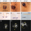
Figure 2
Heart morphogenesis. Dorsal views (AC, G, and H) and cross-sections (DF) of the heart field are shown at three different stages: 23 somites or 20.5 hpf (A,D, and G), 25 somites or 22 hpf (B,E, and H) and 24 hpf (C,F, and I). (AC) Double-labelling in situ hybridization with ventricle myosin heavy chain (vmhc, red) and atrial myosin heavy chain (amhc/myh6, blue) probes. (DF) Cross-sections after in situ hybridization with a cmlc2/myl7 probe (D and E) or a lefty2 probe (blue) with an anti-tropomyosin antibody (brown). (GI) Projections of confocal stacks made from a living Tg(cmlc2:eGFP) embryo. Tracking of individual myocytes was performed, and four examples are shown as yellow dots. After fusion of the bilateral heart fields, a cardiac disc is formed, which is located ventrally to the neural tube and endoderm (A,D, and G). The future ventricle cells (blue cells in A and B) are located in the central region of the disc, where they start to form a cone due to an involution of the tissue at the right side (dashed arrows in B and E). The future atrial cells (blue in A and B) are located at the periphery of the disc. These cells are displaced towards the anterior left and cause the future venous pole to rotate in a clockwise direction (arrows in B, H, and I). Owing to the involution process and rotation processes, the left cardiac field will form the dorsal wall of the heart tube, indicated by lefty2 expression in F. The myocytes derived from the right cardiac field will form the ventral wall of the cardiac tube (lefty-negative cells in F). When the myocardial tube has formed, it will extent anteriorly by a thus far unidentified process.

References

    1. Stainier DY, Lee RK, Fishman MC. Cardiovascular development in the zebrafish. I. Myocardial fate map and heart tube formation. Development. 1993;119:31–40. - PubMed
    1. Keegan BR, Meyer D, Yelon D. Organization of cardiac chamber progenitors in the zebrafish blastula. Development. 2004;131:3081–3091. doi:10.1242/dev.01185. - DOI - PubMed
    1. Keegan BR, Feldman JL, Begemann G, Ingham PW, Yelon D. Retinoic acid signaling restricts the cardiac progenitor pool. Science. 2005;307:247–249. doi:10.1126/science.1101573. - DOI - PubMed
    1. Kishimoto Y, Lee KH, Zon L, Hammerschmidt M, Schulte-Merker S. The molecular nature of zebrafish swirl: BMP2 function is essential during early dorsoventral patterning. Development. 1997;124:4457–4466. - PubMed
    1. Prall OWJ, Menon MK, Solloway MJ, Watanabe Y, Zaffran S, Bajolle F, et al. An Nkx2-5/Bmp2/Smad1 negative feedback loop controls heart progenitor specification and proliferation. Cell. 2007;128:947–959. doi:10.1016/j.cell.2007.01.042. - DOI - PMC - PubMed

Publication types