. 2011 Aug;193(15):3879-86.
doi: 10.1128/JB.00311-11. Epub 2011 May 20.

Broad-spectrum biofilm inhibition by Kingella kingae exopolysaccharide

Affiliations

Broad-spectrum biofilm inhibition by Kingella kingae exopolysaccharide

Meriem Bendaoud et al. J Bacteriol. 2011 Aug.

Abstract

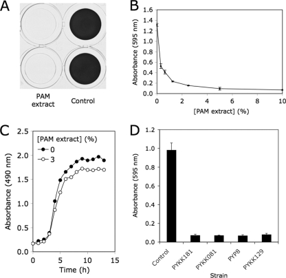
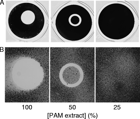
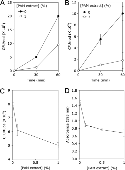
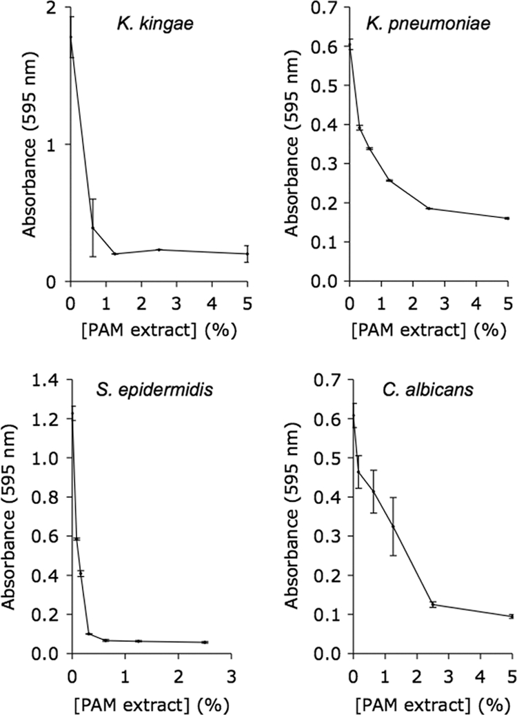
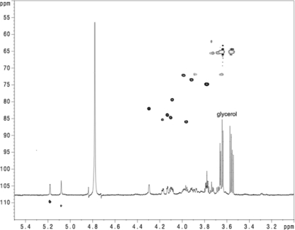
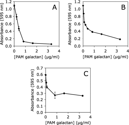
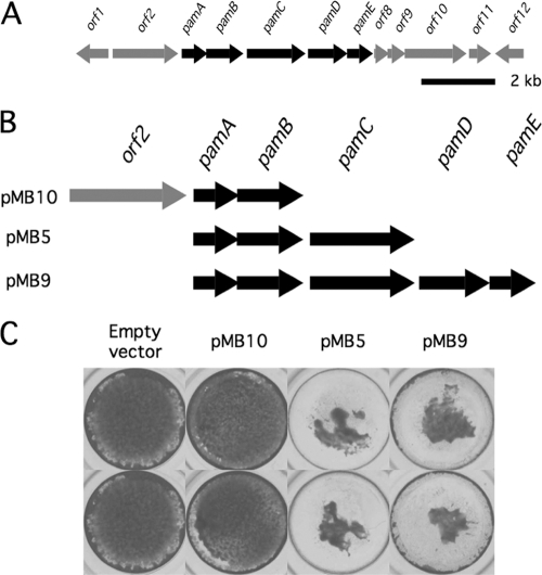
Cell-free extracts prepared from Kingella kingae colony biofilms were found to inhibit biofilm formation by Aggregatibacter actinomycetemcomitans, Klebsiella pneumoniae, Staphylococcus aureus, Staphylococcus epidermidis, Candida albicans, and K. kingae. The extracts evidently inhibited biofilm formation by modifying the physicochemical properties of the cell surface, the biofilm matrix, and the substrate. Chemical and biochemical analyses indicated that the biofilm inhibition activity in the K. kingae extract was due to polysaccharide. Structural analyses showed that the extract contained two major polysaccharides. One was a linear polysaccharide with the structure →6)-α-d-GlcNAcp-(1→5)-β-d-OclAp-(2→, which was identical to a capsular polysaccharide produced by Actinobacillus pleuropneumoniae serotype 5. The second was a novel linear polysaccharide, designated PAM galactan, with the structure →3)-β-d-Galf-(1→6)-β-d-Galf-(1→. Purified PAM galactan exhibited broad-spectrum biofilm inhibition activity. A cluster of three K. kingae genes encoding UDP-galactopyranose mutase (ugm) and two putative galactofuranosyl transferases was sufficient for the synthesis of PAM galactan in Escherichia coli. PAM galactan is one of a growing number of bacterial polysaccharides that exhibit antibiofilm activity. The biological roles and potential technological applications of these molecules remain unknown.


Figures

Fig. 1.
Fig. 1.
Inhibition of S. aureus biofilm formation by K. kingae PAM extract. (A) Biofilm formation by S. aureus SH1000 in 96-well microtiter plate wells in the presence or absence of 10% PAM extract. Biofilms were stained with crystal violet. Duplicate wells are shown. (B) Biofilm formation by S. aureus in the presence of increasing concentrations of PAM extract. Biofilm biomass was quantitated by destaining the biofilm and measuring the absorbance of the crystal violet solution. Values show mean absorbance and range for duplicate wells. (C) Growth of S. aureus strain SH1000 in the presence or absence of 3% PAM extract. Growth was monitored by measuring the absorbance of the culture at 490 nm. Values show mean absorbance for duplicate tubes. Error bars were omitted for clarity. (D) Inhibition of S. aureus SH1000 biofilm formation by colony biofilm extracts prepared from four K. kingae clinical strains. Extracts were tested at a concentration of 10% by volume. Values show mean absorbance and range for duplicate wells.
Fig. 2.
Fig. 2.
Polystyrene surfaces coated with PAM extract inhibit biofilm formation. Drops of PAM extract were pipetted onto the surface of a 24-well microtiter plate and allowed to evaporate. In some wells the PAM extract was diluted with 1 or 3 volumes of water. Wells were then inoculated with S. epidermidis (A) or A. actinomycetemcomitans (B) and incubated at 37°C. After 18 h, wells were rinsed with water and stained with crystal violet.
Fig. 3.
Fig. 3.
PAM extract inhibits S. aureus surface attachment and disperses preformed S. aureus biofilms. (A and B) Binding of S. aureus cells to stainless steel (A) and polystyrene (B) rods in the presence or absence of 3% PAM extract. Values show mean CFU per rod and range for duplicate rods. (C) Binding of S. aureus cells to polycarbonate tubes in 15 min in the presence of 0, 0.1, or 1% PAM extract. Values show mean CFU per tube and range for duplicate tubes. (D) Detachment of 18-h-old S. aureus biofilms by PAM extract. Biofilms were rinsed with water and treated with the indicated concentration of PAM extract for 1 h. Biofilms were then rinsed with water and quantitated by crystal violet staining. Values show mean absorbance and range for duplicate wells.
Fig. 4.
Fig. 4.
PAM extract inhibits biofilm formation by K. kingae, K. pneumoniae, S. epidermidis, and C. albicans. Biofilms were cultured in 96-well microtiter plates and stained with crystal violet. Values shown mean absorbance and range for duplicate wells.
Fig. 5.
Fig. 5.
1H-13C heteronuclear multiple-quantum coherence (HMQC) spectrum of PAM extract from K. kingae strain PYKK181.
Fig. 6.
Fig. 6.
Inhibition of S. aureus SH1000 (A), K. kingae PYKK181 (B), and K. pneumoniae 1840 (C) biofilm formation by purified K. kingae PAM galactan. Biofilm biomass was quantitated by crystal violet staining. Graphs show mean crystal violet absorbance values and range for duplicate wells.
Fig. 7.
Fig. 7.
Expression of the K. kingae pam genes in E. coli. (A) Genetic map of the K. kingae pam genes and surrounding region. Arrows indicate open reading frames and direction of transcription. Scale bar, 2 kb. (B) Genetic maps of expression plasmid inserts. (C) Inhibition of S. aureus biofilm formation by colony biofilm extracts isolated from plasmid-harboring E. coli strains. Biofilms were stained with crystal violet and photographed. Duplicate wells are shown.

References

    1. Altman E., Brisson J.-R., Perry M. B. 1987. Structure of the capsular polysaccharide of Haemophilus pleuropneumoniae serotype 5. Eur. J. Biochem. 170:185–192 - PubMed
    1. Berbari E. F., Cockerill F. R., Stickelberg J. M. 1997. Infective endocarditis due to unusual or fastidious microorganisms. Mayo Clin. Proc. 72:532–542 - PubMed
    1. Bock K., Pedersen C. 1983. Carbon-13 nuclear magnetic resonance spectroscopy of monosaccharides. Adv. Carbohydr. Chem. Biochem. 41:27–66
    1. Byrd M. S., et al. 2009. Genetic and biochemical analyses of the Pseudomonas aeruginosa Psl exopolysaccharide reveal overlapping roles for polysaccharide synthesis enzymes in Psl and LPS production. Mol. Microbiol. 73:622–638 - PMC - PubMed
    1. Colvin K. M., et al. 2011. The Pel polysaccharide can serve a structural and protective role in the biofilm matrix of Pseudomonas aeruginosa. PLoS Pathog. 7:e1001264. - PMC - PubMed

Publication types

MeSH terms

Substances

Associated data

LinkOut - more resources